The Heart Research Institute, Sydney, NSW, 2042, Australia.
Sydney Medical School, University of Sydney, Sydney, NSW, 2006, Australia.
Stem Cell Res Ther. 2018 Mar 21;9(1):70. doi: 10.1186/s13287-018-0824-2.
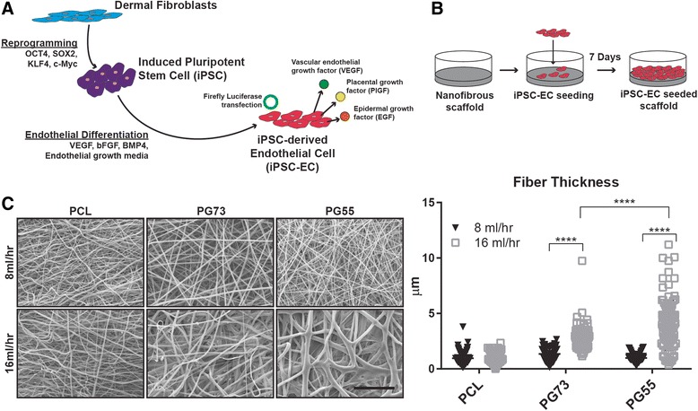
Induced pluripotent stem-cell derived endothelial cells (iPSC-ECs) can be generated from any somatic cell and their iPSC sources possess unlimited self-renewal. Previous demonstration of their proangiogenic activity makes them a promising cell type for treatment of ischemic injury. As with many other stem cell approaches, the low rate of in-vivo survival has been a major limitation to the efficacy of iPSC-ECs to date. In this study, we aimed to increase the in-vivo lifetime of iPSC-ECs by culturing them on electrospun polycaprolactone (PCL)/gelatin scaffolds, before quantifying the subsequent impact on their proangiogenic function.
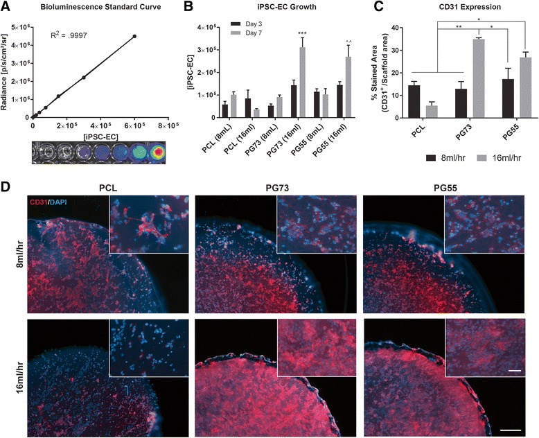
iPSC-ECs were isolated and stably transfected with a luciferase reporter to facilitate quantification of cell numbers and non-invasive imaging in-vivo PCL/gelatin scaffolds were engineered using electrospinning to obtain woven meshes of nanofibers. iPSC-ECs were cultured on scaffolds for 7 days. Subsequently, cell growth and function were assessed in vitro followed by implantation in a mouseback subcutaneous model for 7 days.
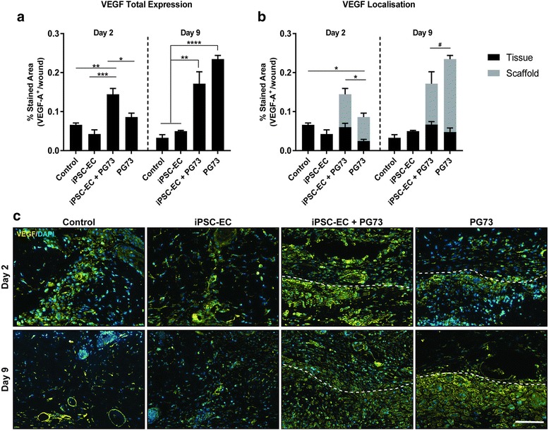
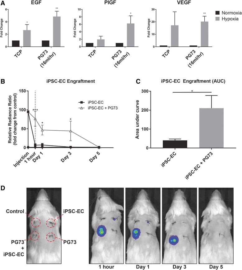
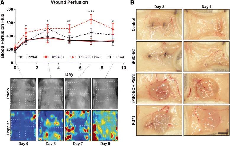
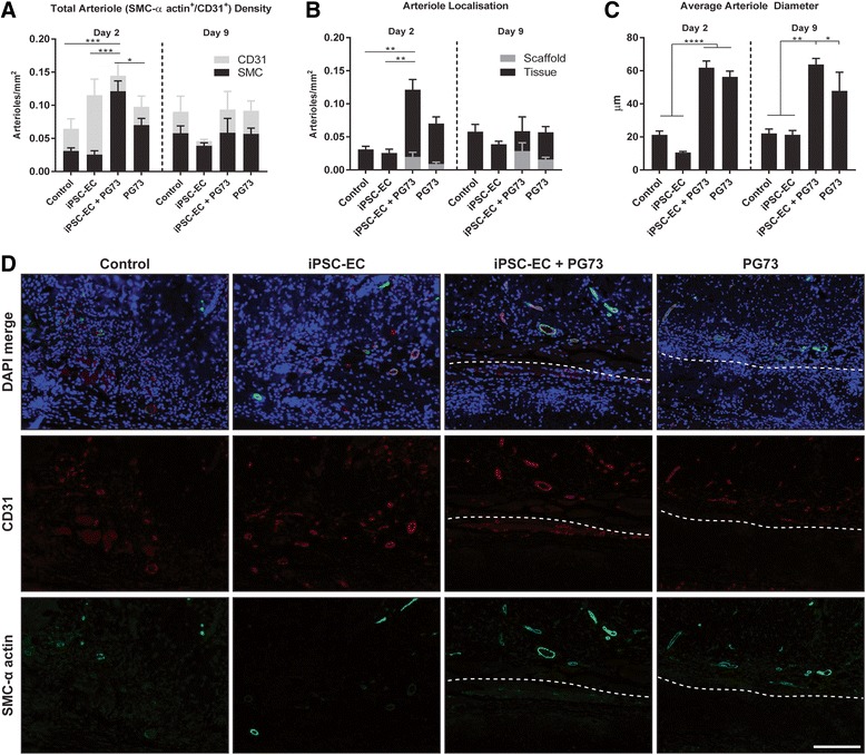
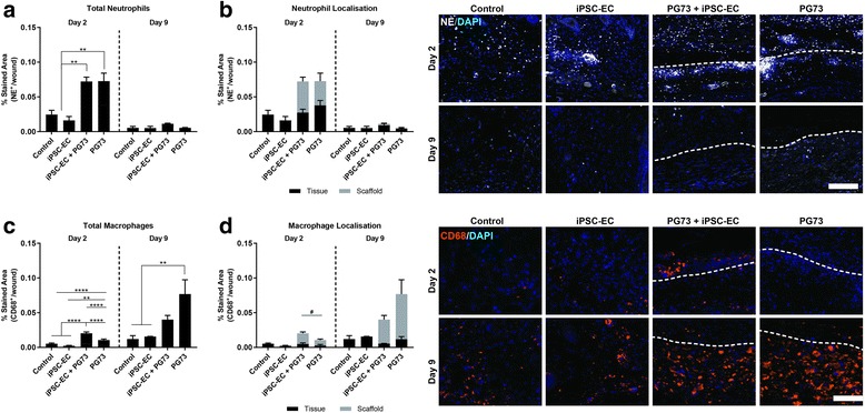
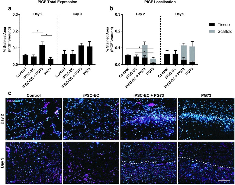
Using a matrix of conditions, we found that scaffold blends with ratios of PCL:gelatin of 70:30 (PG73) spun at high flow rates supported the greatest levels of iPSC-EC growth, retention of phenotype, and function in vitro. Implanting iPSC-ECs seeded on PG73 scaffolds in vivo improved their survival up to 3 days, compared to cells directly injected into control wounds, which were no longer observable within 1 h. Enhanced engraftment improved blood perfusion, observed through non-invasive laser Doppler imaging. Immunohistochemistry revealed a corresponding increase in host angiogenic mechanisms characterized by the enhanced recruitment of macrophages and the elevated expression of proangiogenic cytokines vascular endothelial growth factor and placental growth factor.
Knowledge of these mechanisms combined with a deeper understanding of the scaffold parameters influencing this function provides the groundwork for optimizing future iPSC-EC therapies utilizing engraftment platforms. The development of combined scaffold and iPSC-EC therapies could ultimately improve therapeutic angiogenesis and the treatment of ischemic injury.
诱导多能干细胞衍生的内皮细胞(iPSC-ECs)可由任何体细胞生成,其 iPSC 来源具有无限的自我更新能力。先前的研究表明它们具有促血管生成活性,这使它们成为治疗缺血性损伤的有前途的细胞类型。与许多其他干细胞方法一样,iPSC-ECs 目前在体内的存活率低是其疗效的主要限制。在这项研究中,我们旨在通过在静电纺丝聚己内酯(PCL)/明胶支架上培养 iPSC-ECs 来增加其在体内的寿命,然后定量评估其对促血管生成功能的后续影响。
分离 iPSC-ECs 并稳定转染荧光素酶报告基因,以方便在体内进行细胞数量的定量和非侵入性成像。使用静电纺丝工程设计 PCL/明胶支架,以获得纳米纤维的编织网格。将 iPSC-ECs 在支架上培养 7 天。随后,在体外评估细胞生长和功能,然后将其植入小鼠背部皮下模型中 7 天。
通过一系列条件,我们发现以 70:30(PG73)的比例混合的支架在高流速下纺丝时,最支持 iPSC-EC 的生长、体外保留表型和功能。与直接注射到对照伤口中的细胞相比,将接种在 PG73 支架上的 iPSC-ECs 植入体内可将其存活率提高至 3 天,而对照伤口中的细胞在 1 小时内不再可见。增强的植入可通过非侵入性激光多普勒成像观察到改善的血液灌注。免疫组织化学显示,宿主血管生成机制得到相应增强,表现为巨噬细胞的募集增强和促血管生成细胞因子血管内皮生长因子和胎盘生长因子的表达升高。
这些机制的知识以及对影响该功能的支架参数的更深入理解为优化利用植入平台的未来 iPSC-EC 治疗提供了基础。联合支架和 iPSC-EC 治疗的发展最终可能会改善治疗性血管生成和缺血性损伤的治疗。