• 文献检索
  • 文档翻译
  • 深度研究
  • 学术资讯
  • Suppr Zotero 插件Zotero 插件
  • 邀请有礼
  • 套餐&价格
  • 历史记录
应用&插件
Suppr Zotero 插件Zotero 插件浏览器插件Mac 客户端Windows 客户端微信小程序
定价
高级版会员购买积分包购买API积分包
服务
文献检索文档翻译深度研究API 文档MCP 服务
关于我们
关于 Suppr公司介绍联系我们用户协议隐私条款
关注我们

Suppr 超能文献

核心技术专利:CN118964589B侵权必究
粤ICP备2023148730 号-1Suppr @ 2026

文献检索

告别复杂PubMed语法,用中文像聊天一样搜索,搜遍4000万医学文献。AI智能推荐,让科研检索更轻松。

立即免费搜索

文件翻译

保留排版,准确专业,支持PDF/Word/PPT等文件格式,支持 12+语言互译。

免费翻译文档

深度研究

AI帮你快速写综述,25分钟生成高质量综述,智能提取关键信息,辅助科研写作。

立即免费体验

基于青光眼性损伤的视网膜神经纤维束正面图像定制视野检查位置

Customizing Perimetric Locations Based on En Face Images of Retinal Nerve Fiber Bundles With Glaucomatous Damage.

作者信息

Alluwimi Muhammed S, Swanson William H, Malinovsky Victor E, King Brett J

机构信息

Indiana University School of Optometry, Bloomington, IN, USA.

Qassim University Department of Optometry, College of Applied Medical Sciences, Qassim, Saudi Arabia.

出版信息

Transl Vis Sci Technol. 2018 Mar 15;7(2):5. doi: 10.1167/tvst.7.2.5. eCollection 2018 Mar.

DOI:10.1167/tvst.7.2.5
PMID:29576929
原文链接:https://pmc.ncbi.nlm.nih.gov/articles/PMC5858465/
Abstract

PURPOSE

Prior studies suggested the use of customized perimetric locations in glaucoma; these studies were limited by imaging only the superficial depths of the retinal nerve fiber layer (RNFL) and by prolonged perimetric testing. We aimed to develop a rapid perimetric test guided by high-resolution images of RNFL bundles.

METHODS

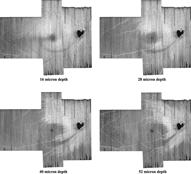
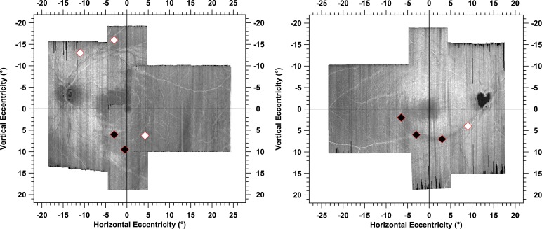
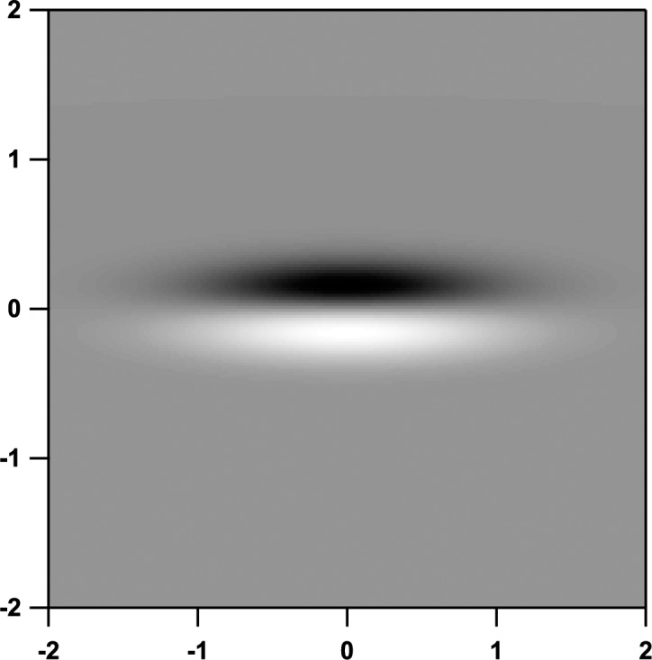
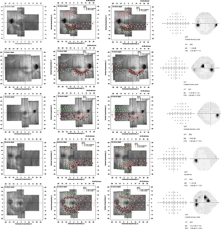
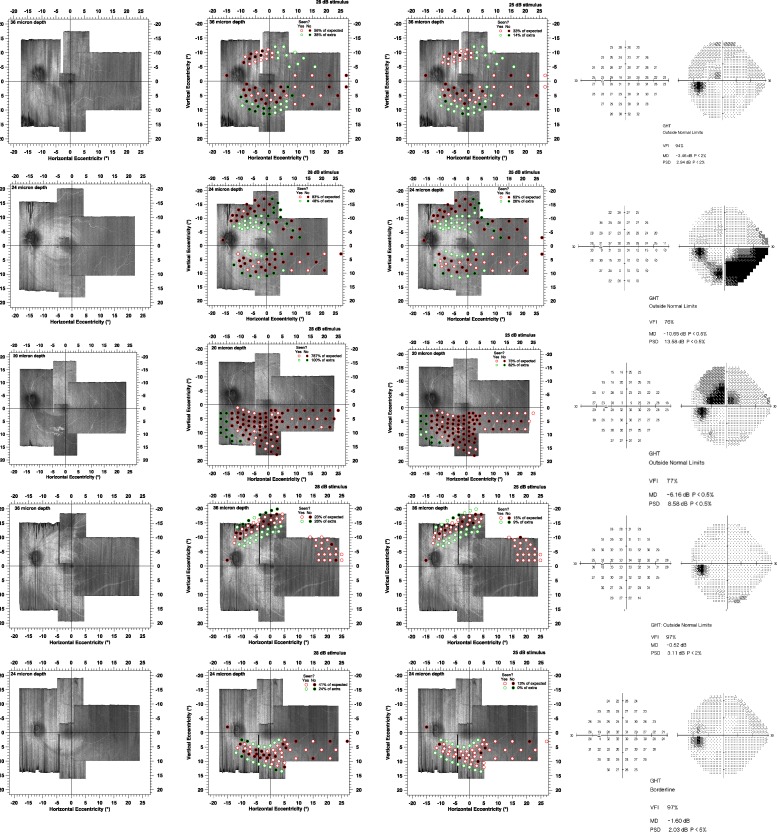
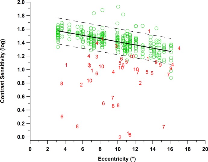
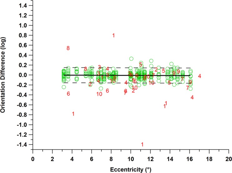
We recruited 10 patients with glaucoma, ages 56 to 80 years, median 68 years, and 10 controls, ages 55 to 77 years, median 68 years. The patients were selected based on discrepancies between locations of glaucomatous damage for perimetric and structural measures. Montaging was used to produce optical coherence tomography en face images of the RNFL covering much of the 24-2 grid locations. In experiment 1, we presented the Goldmann size III stimulus at preselected retinal locations of glaucomatous damage, using just two contrasts. In experiment 2, we developed an elongated sinusoidal stimulus, aligned within the defect, to measure contrast sensitivities; abnormalities were defined based on lower 95% reference limits derived from the controls.

RESULTS

The percentage of predicted locations where size III was not seen at 28 dB ranged from 16% to 80%, with a median of 48%. Contrast sensitivity for the sinusoidal stimulus was below the 95% reference range for 37 of 44 stimuli aligned within the defects.

CONCLUSIONS

We developed methods for rapid perimetric testing guided by en face images of the RNFL bundles in patients with glaucoma. Results indicated ganglion cell damage under all of the visible RNFL defects.

TRANSLATIONAL RELEVANCE

Customized perimetric locations have potential to improve clinical assessment of glaucoma.

摘要

目的

先前的研究表明在青光眼患者中可使用定制的视野检查位置;这些研究存在局限性,仅对视网膜神经纤维层(RNFL)的浅表深度进行成像,且视野检查时间较长。我们旨在开发一种由RNFL束的高分辨率图像引导的快速视野检查方法。

方法

我们招募了10例青光眼患者,年龄在56至80岁之间,中位数为68岁,以及10名对照者,年龄在55至77岁之间,中位数为68岁。根据视野检查和结构测量中青光眼损伤位置的差异选择患者。采用拼接技术生成覆盖24-2网格位置大部分区域的RNFL正面光学相干断层扫描图像。在实验1中,我们仅使用两种对比度,在预先选定的青光眼损伤视网膜位置呈现戈德曼III号视标。在实验2中,我们开发了一种在缺损内对齐的细长正弦波视标,以测量对比度敏感度;根据对照组得出的较低95%参考限值来定义异常情况。

结果

在28 dB时未看到III号视标的预测位置百分比范围为16%至80%,中位数为48%。在缺损内对齐的44个视标中,有37个正弦波视标的对比度敏感度低于95%参考范围。

结论

我们开发了由青光眼患者RNFL束正面图像引导的快速视野检查方法。结果表明在所有可见的RNFL缺损下方均存在神经节细胞损伤。

转化相关性

定制的视野检查位置有可能改善青光眼的临床评估。

https://cdn.ncbi.nlm.nih.gov/pmc/blobs/f5a7/5858465/11a413c6e3c5/i2164-2591-7-2-5-f07.jpg
https://cdn.ncbi.nlm.nih.gov/pmc/blobs/f5a7/5858465/41ba72bcfee5/i2164-2591-7-2-5-f01.jpg
https://cdn.ncbi.nlm.nih.gov/pmc/blobs/f5a7/5858465/8f7e30a3bc5c/i2164-2591-7-2-5-f02.jpg
https://cdn.ncbi.nlm.nih.gov/pmc/blobs/f5a7/5858465/2d7be930edaa/i2164-2591-7-2-5-f03.jpg
https://cdn.ncbi.nlm.nih.gov/pmc/blobs/f5a7/5858465/aa427d4e71b1/i2164-2591-7-2-5-f04.jpg
https://cdn.ncbi.nlm.nih.gov/pmc/blobs/f5a7/5858465/f569022ff4a9/i2164-2591-7-2-5-f05.jpg
https://cdn.ncbi.nlm.nih.gov/pmc/blobs/f5a7/5858465/58b1dc2dae8f/i2164-2591-7-2-5-f06.jpg
https://cdn.ncbi.nlm.nih.gov/pmc/blobs/f5a7/5858465/11a413c6e3c5/i2164-2591-7-2-5-f07.jpg

相似文献

1
Customizing Perimetric Locations Based on En Face Images of Retinal Nerve Fiber Bundles With Glaucomatous Damage.基于青光眼性损伤的视网膜神经纤维束正面图像定制视野检查位置
Transl Vis Sci Technol. 2018 Mar 15;7(2):5. doi: 10.1167/tvst.7.2.5. eCollection 2018 Mar.
2
A basis for customising perimetric locations within the macula in glaucoma.青光眼黄斑区内视野检查位置定制的基础。
Ophthalmic Physiol Opt. 2018 Mar;38(2):164-173. doi: 10.1111/opo.12435. Epub 2018 Jan 8.
3
Structure-function assessment in glaucoma based on perimetric sensitivity and en face optical coherence tomography images of retinal nerve fiber bundles.基于视野敏感性和视网膜神经纤维束的共焦光学相干断层扫描图像的青光眼结构-功能评估。
Sci Rep. 2023 Feb 13;13(1):2497. doi: 10.1038/s41598-023-28917-1.
4
A Novel Stimulus to Improve Perimetric Sampling within the Macula in Patients with Glaucoma.一种改善青光眼患者黄斑区内视野取样的新刺激方法。
Optom Vis Sci. 2021 Apr 1;98(4):374-383. doi: 10.1097/OPX.0000000000001677.
5
Comparison of defect depths for sinusoidal and circular perimetric stimuli in patients with glaucoma.青光眼患者中正弦和圆形周边视野刺激的缺损深度比较。
Ophthalmic Physiol Opt. 2019 Jan;39(1):26-36. doi: 10.1111/opo.12598.
6
Functional characteristics of glaucoma related arcuate defects seen on OCT en face visualisation of the retinal nerve fibre layer.OCT 眼前节断层成像对视神经纤维层弓形缺损的青光眼相关功能特征。
Ophthalmic Physiol Opt. 2021 Mar;41(2):437-446. doi: 10.1111/opo.12780. Epub 2021 Jan 25.
7
Localized Retinal Nerve Fiber Layer Defects in Red-free Photographs Versus En Face Structural Optical Coherence Tomography Images.眼底自发荧光与共焦激光扫描检眼镜检查在眼挫伤诊断中的比较
J Glaucoma. 2018 Mar;27(3):269-274. doi: 10.1097/IJG.0000000000000866.
8
Localized Retinal Nerve Fiber Layer Defect Location Among Red-free Fundus Photographs, En Face Structural Images, and Cirrus HD-OCT Maps.眼底自发荧光图像、共焦激光扫描检眼镜结构像和 Cirrus HD-OCT 断层扫描图中红色滤光片下局限性视网膜神经纤维层缺损定位。
J Glaucoma. 2019 Dec;28(12):1054-1060. doi: 10.1097/IJG.0000000000001388.
9
Assessment of the reliability of standard automated perimetry in regions of glaucomatous damage.评估标准自动视野计在青光眼损害区域的可靠性。
Ophthalmology. 2014 Jul;121(7):1359-69. doi: 10.1016/j.ophtha.2014.01.020. Epub 2014 Mar 12.
10
Novel Technique for Quantifying Retinal Nerve Fiber Bundle Abnormality in the Temporal Raphe.量化颞侧中缝处视网膜神经纤维束异常的新技术。
Optom Vis Sci. 2018 Apr;95(4):309-317. doi: 10.1097/OPX.0000000000001202.

引用本文的文献

1
Predicting perimetric defects from en face maps of retinal nerve fibre layer reflectance.从视网膜神经纤维层反射的正面图预测周边视野缺损。
Ophthalmic Physiol Opt. 2024 May;44(3):613-625. doi: 10.1111/opo.13289. Epub 2024 Feb 25.
2
Structure-function relationship in glaucoma: Optical coherence tomography en face imaging vs. red-free fundus photography.青光眼的结构-功能关系:光学相干断层扫描(OCT)眼前节成像与无赤光眼底照相。
Eye (Lond). 2023 Oct;37(14):2969-2976. doi: 10.1038/s41433-023-02452-9. Epub 2023 Feb 22.
3
Structure-function assessment in glaucoma based on perimetric sensitivity and en face optical coherence tomography images of retinal nerve fiber bundles.

本文引用的文献

1
Individual differences in the shape of the nasal visual field.鼻侧视野形状的个体差异。
Vision Res. 2017 Dec;141:23-29. doi: 10.1016/j.visres.2016.04.001. Epub 2016 Jun 16.
2
Between-Subject Variability in Healthy Eyes as a Primary Source of Structural-Functional Discordance in Patients With Glaucoma.健康眼睛的个体间变异性是青光眼患者结构-功能不一致的主要来源。
Invest Ophthalmol Vis Sci. 2016 Feb;57(2):502-7. doi: 10.1167/iovs.15-18633.
3
Choice of Stimulus Range and Size Can Reduce Test-Retest Variability in Glaucomatous Visual Field Defects.
基于视野敏感性和视网膜神经纤维束的共焦光学相干断层扫描图像的青光眼结构-功能评估。
Sci Rep. 2023 Feb 13;13(1):2497. doi: 10.1038/s41598-023-28917-1.
4
Within-eye and between-subject variability for reflectance of the retinal nerve fibre layer.眼内和个体间视网膜神经纤维层反射率的可变性。
Ophthalmic Physiol Opt. 2022 Nov;42(6):1316-1325. doi: 10.1111/opo.13027. Epub 2022 Aug 1.
5
Enhanced Objective Detection of Retinal Nerve Fiber Bundle Defects in Glaucoma With a Novel Method for En Face OCT Slab Image Construction and Analysis.利用一种新的 OCT 切片图像构建和分析方法增强青光眼视网膜神经纤维束缺陷的客观检测。
Transl Vis Sci Technol. 2021 Oct 4;10(12):1. doi: 10.1167/tvst.10.12.1.
6
A Simple Subjective Evaluation of Enface OCT Reflectance Images Distinguishes Glaucoma From Healthy Eyes.正视 OCT 反射图像的简单主观评估可区分青光眼与健康眼。
Transl Vis Sci Technol. 2021 May 3;10(6):31. doi: 10.1167/tvst.10.6.31.
7
Functional characteristics of glaucoma related arcuate defects seen on OCT en face visualisation of the retinal nerve fibre layer.OCT 眼前节断层成像对视神经纤维层弓形缺损的青光眼相关功能特征。
Ophthalmic Physiol Opt. 2021 Mar;41(2):437-446. doi: 10.1111/opo.12780. Epub 2021 Jan 25.
8
The Relationship Between Macula Retinal Ganglion Cell Density and Visual Function in the Nonhuman Primate.灵长类动物的黄斑视网膜神经节细胞密度与视觉功能的关系。
Invest Ophthalmol Vis Sci. 2021 Jan 4;62(1):5. doi: 10.1167/iovs.62.1.5.
9
Presumed activated retinal astrocytes and Müller cells in healthy and glaucomatous eyes detected by spectral domain optical coherence tomography.通过谱域光相干断层扫描检测健康眼和青光眼眼中假定的激活的视网膜星形胶质细胞和 Müller 细胞。
Ophthalmic Physiol Opt. 2020 Nov;40(6):738-751. doi: 10.1111/opo.12731. Epub 2020 Sep 4.
10
Estimation of the central 10-degree visual field using en-face images obtained by optical coherence tomography.利用光学相干断层扫描获得的共焦图像估计中央 10 度视野。
PLoS One. 2020 Mar 5;15(3):e0229867. doi: 10.1371/journal.pone.0229867. eCollection 2020.
刺激范围和大小的选择可降低青光眼视野缺损的重测变异性。
Transl Vis Sci Technol. 2014 Sep 25;3(5):6. doi: 10.1167/tvst.3.5.6. eCollection 2014 Sep.
4
Contrast sensitivity perimetry and clinical measures of glaucomatous damage.青光眼性损害的对比敏感度视野检查与临床测量
Optom Vis Sci. 2014 Nov;91(11):1302-11. doi: 10.1097/OPX.0000000000000395.
5
Imaging of the temporal raphe with optical coherence tomography.使用光学相干断层扫描对中缝颞核进行成像。
Ophthalmology. 2014 Nov;121(11):2287-8. doi: 10.1016/j.ophtha.2014.06.023. Epub 2014 Aug 22.
6
In vivo adaptive optics imaging of the temporal raphe and its relationship to the optic disc and fovea in the human retina.人视网膜中颞侧中缝的体内自适应光学成像及其与视盘和中央凹的关系。
Invest Ophthalmol Vis Sci. 2014 Aug 21;55(9):5952-61. doi: 10.1167/iovs.14-14893.
7
A Test of a Model of Glaucomatous Damage of the Macula With High-Density Perimetry: Implications for the Locations of Visual Field Test Points.用高密度视野计对黄斑部青光眼性损害模型的测试:对视野测试点位置的启示
Transl Vis Sci Technol. 2014 Jun 19;3(3):5. doi: 10.1167/tvst.3.3.5. eCollection 2014 May.
8
A novel strategy for the estimation of the general height of the visual field in patients with glaucoma.一种用于估计青光眼患者总体视野高度的新策略。
Graefes Arch Clin Exp Ophthalmol. 2014 May;252(5):801-9. doi: 10.1007/s00417-014-2602-x. Epub 2014 Mar 18.
9
Structure-function mapping: variability and conviction in tracing retinal nerve fiber bundles and comparison to a computational model.结构-功能映射:追踪视网膜神经纤维束的变异性和可信度,以及与计算模型的比较。
Invest Ophthalmol Vis Sci. 2014 Feb 4;55(2):728-36. doi: 10.1167/iovs.13-13142.
10
The influence of intersubject variability in ocular anatomical variables on the mapping of retinal locations to the retinal nerve fiber layer and optic nerve head.眼解剖学变量的个体间变异性对视网膜位置到视网膜神经纤维层和视盘映射的影响。
Invest Ophthalmol Vis Sci. 2013 Sep 9;54(9):6074-82. doi: 10.1167/iovs.13-11902.