The Krasnow Institute for Advanced Study, George Mason University, Fairfax, VA, USA.
Department of Bioengineering, George Mason University, Fairfax, VA, USA.
Eur J Neurosci. 2019 Mar;49(6):768-783. doi: 10.1111/ejn.13919. Epub 2018 Apr 20.
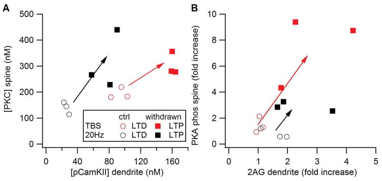
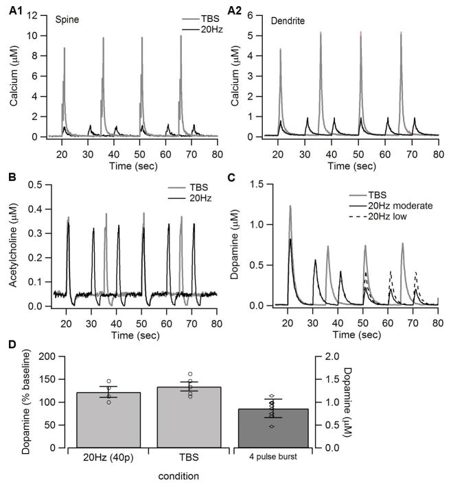
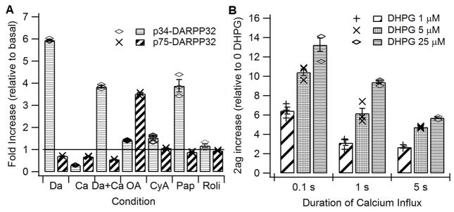
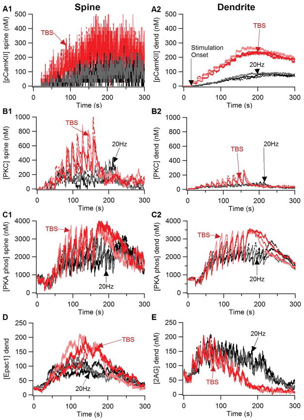
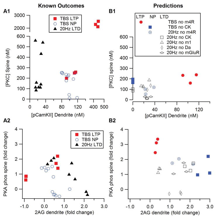
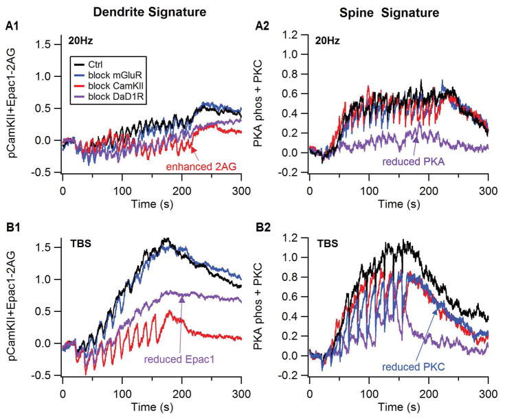
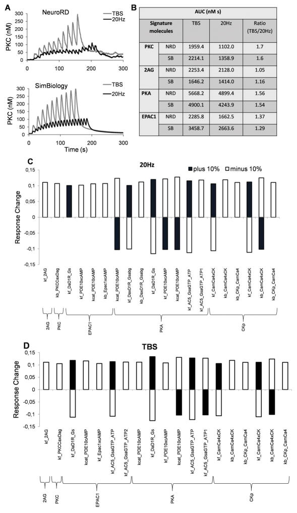
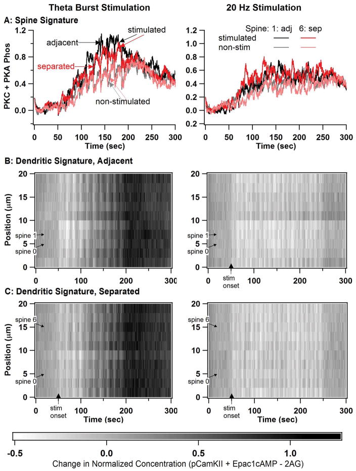
The striatum, the input structure of the basal ganglia, is a major site of learning and memory for goal-directed actions and habit formation. Spiny projection neurons of the striatum integrate cortical, thalamic, and nigral inputs to learn associations, with cortico-striatal synaptic plasticity as a learning mechanism. Signaling molecules implicated in synaptic plasticity are altered in alcohol withdrawal, which may contribute to overly strong learning and increased alcohol seeking and consumption. To understand how interactions among signaling molecules produce synaptic plasticity, we implemented a mechanistic model of signaling pathways activated by dopamine D1 receptors, acetylcholine receptors, and glutamate. We use our novel, computationally efficient simulator, NeuroRD, to simulate stochastic interactions both within and between dendritic spines. Dopamine release during theta burst and 20-Hz stimulation was extrapolated from fast-scan cyclic voltammetry data collected in mouse striatal slices. Our results show that the combined activity of several key plasticity molecules correctly predicts the occurrence of either LTP, LTD, or no plasticity for numerous experimental protocols. To investigate spatial interactions, we stimulate two spines, either adjacent or separated on a 20-μm dendritic segment. Our results show that molecules underlying LTP exhibit spatial specificity, whereas 2-arachidonoylglycerol exhibits a spatially diffuse elevation. We also implement changes in NMDA receptors, adenylyl cyclase, and G protein signaling that have been measured following chronic alcohol treatment. Simulations under these conditions suggest that the molecular changes can predict changes in synaptic plasticity, thereby accounting for some aspects of alcohol use disorder.
纹状体是基底神经节的输入结构,是学习和记忆目标导向动作和习惯形成的主要部位。纹状体的棘突投射神经元整合皮质、丘脑和黑质的输入以学习关联,皮质-纹状体突触可塑性是一种学习机制。在酒精戒断期间,涉及突触可塑性的信号分子发生改变,这可能导致过度强烈的学习以及增加的酒精寻求和消费。为了了解信号分子之间的相互作用如何产生突触可塑性,我们实施了一种由多巴胺 D1 受体、乙酰胆碱受体和谷氨酸激活的信号通路的机制模型。我们使用我们的新型、计算效率高的模拟器 NeuroRD 来模拟树突棘内和树突棘之间的随机相互作用。在从在小鼠纹状体切片中收集的快速扫描循环伏安法数据中推断出 theta 爆发和 20-Hz 刺激期间的多巴胺释放。我们的结果表明,几个关键可塑性分子的组合活性正确预测了许多实验方案中 LTP、 LTD 或无可塑性的发生。为了研究空间相互作用,我们刺激两个树突棘,要么相邻,要么在 20μm 的树突段上分开。我们的结果表明,LTP 下的分子表现出空间特异性,而 2-花生四烯酸甘油则表现出空间弥散性升高。我们还实施了 NMDA 受体、腺苷酸环化酶和 G 蛋白信号的变化,这些变化是在慢性酒精处理后测量的。在这些条件下的模拟表明,分子变化可以预测突触可塑性的变化,从而解释了一些酒精使用障碍的方面。