Miguel-Hidalgo José J
Department of Psychiatry and Human Behavior, University of Mississippi Medical Center, Jackson, MS, United States.
Front Mol Neurosci. 2018 Mar 20;11:78. doi: 10.3389/fnmol.2018.00078. eCollection 2018.
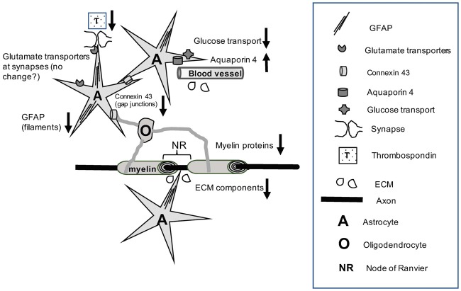
Postmortem studies reveal structural and molecular alterations of astrocytes and oligodendrocytes in both the gray and white matter (GM and WM) of the prefrontal cortex (PFC) in human subjects with chronic alcohol abuse or dependence. These glial cellular changes appear to parallel and may largely explain structural and functional alterations detected using neuroimaging techniques in subjects with alcohol use disorders (AUDs). Moreover, due to the crucial roles of astrocytes and oligodendrocytes in neurotransmission and signal conduction, these cells are very likely major players in the molecular mechanisms underpinning alcoholism-related connectivity disturbances between the PFC and relevant interconnecting brain regions. The glia-mediated etiology of alcohol-related brain damage is likely multifactorial since metabolic, hormonal, hepatic and hemodynamic factors as well as direct actions of ethanol or its metabolites have the potential to disrupt distinct aspects of glial neurobiology. Studies in animal models of alcoholism and postmortem human brains have identified astrocyte markers altered in response to significant exposures to ethanol or during alcohol withdrawal, such as gap-junction proteins, glutamate transporters or enzymes related to glutamate and gamma-aminobutyric acid (GABA) metabolism. Changes in these proteins and their regulatory pathways would not only cause GM neuronal dysfunction, but also disturbances in the ability of WM axons to convey impulses. In addition, alcoholism alters the expression of astrocyte and myelin proteins and of oligodendrocyte transcription factors important for the maintenance and plasticity of myelin sheaths in WM and GM. These changes are concomitant with epigenetic DNA and histone modifications as well as alterations in regulatory microRNAs (miRNAs) that likely cause profound disturbances of gene expression and protein translation. Knowledge is also available about interactions between astrocytes and oligodendrocytes not only at the Nodes of Ranvier (NR), but also in gap junction-based astrocyte-oligodendrocyte contacts and other forms of cell-to-cell communication now understood to be critical for the maintenance and formation of myelin. Close interactions between astrocytes and oligodendrocytes also suggest that therapies for alcoholism based on a specific glial cell type pathology will require a better understanding of molecular interactions between different cell types, as well as considering the possibility of using combined molecular approaches for more effective therapies.
尸检研究表明,在患有慢性酒精滥用或酒精依赖的人类受试者中,前额叶皮质(PFC)的灰质和白质(GM和WM)中的星形胶质细胞和少突胶质细胞存在结构和分子改变。这些胶质细胞变化似乎与酒精使用障碍(AUDs)患者中使用神经成像技术检测到的结构和功能改变相平行,并且可能在很大程度上解释了这些改变。此外,由于星形胶质细胞和少突胶质细胞在神经传递和信号传导中起关键作用,这些细胞很可能是导致PFC与相关互连脑区之间酒精中毒相关连接障碍的分子机制中的主要参与者。酒精相关脑损伤的胶质细胞介导病因可能是多因素的,因为代谢、激素、肝脏和血流动力学因素以及乙醇或其代谢产物的直接作用有可能破坏胶质神经生物学的不同方面。对酒精中毒动物模型和人类尸检大脑的研究已经确定,在大量接触乙醇或戒酒期间,星形胶质细胞标志物会发生改变,例如缝隙连接蛋白、谷氨酸转运体或与谷氨酸和γ-氨基丁酸(GABA)代谢相关的酶。这些蛋白质及其调节途径的变化不仅会导致GM神经元功能障碍,还会干扰WM轴突传递冲动的能力。此外,酒精中毒会改变星形胶质细胞和髓鞘蛋白以及少突胶质细胞转录因子的表达,这些因子对WM和GM中髓鞘的维持和可塑性很重要。这些变化与表观遗传DNA和组蛋白修饰以及调节性微小RNA(miRNA)的改变同时发生,这些改变可能会导致基因表达和蛋白质翻译的严重紊乱。现在人们也了解到星形胶质细胞和少突胶质细胞之间的相互作用,不仅在郎飞结(NR)处,而且在基于缝隙连接的星形胶质细胞 - 少突胶质细胞接触以及其他形式的细胞间通讯中,这些相互作用现在被认为对髓鞘的维持和形成至关重要。星形胶质细胞和少突胶质细胞之间的密切相互作用还表明,基于特定胶质细胞类型病理学的酒精中毒治疗方法需要更好地理解不同细胞类型之间的分子相互作用,并考虑使用联合分子方法进行更有效治疗的可能性。