Genomics and Epigenetics Division, Garvan Institute of Medical Research, New South Wales 2010, Australia.
St Vincent's Clinical School, University of New South Wales, New South Wales 2052, Australia.
Genome Res. 2018 May;28(5):726-738. doi: 10.1101/gr.227975.117. Epub 2018 Apr 4.
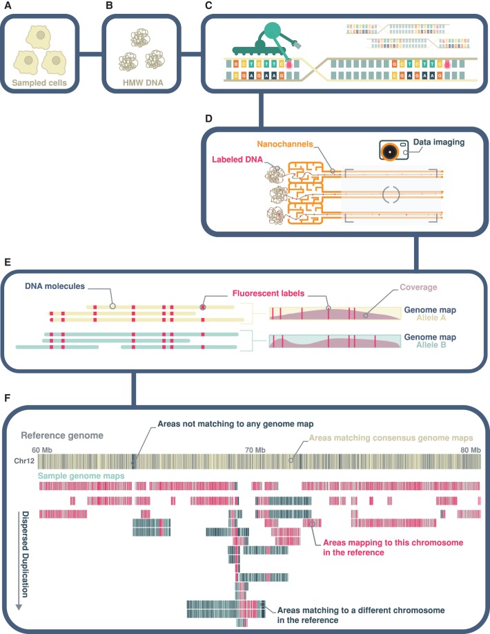
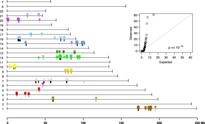
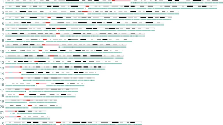
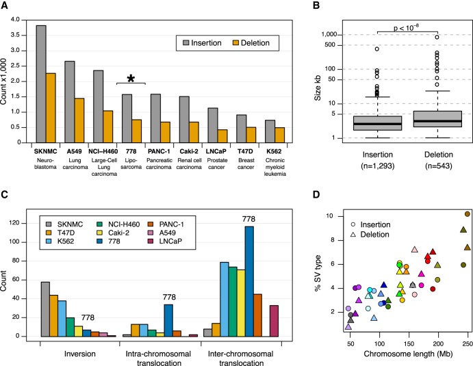
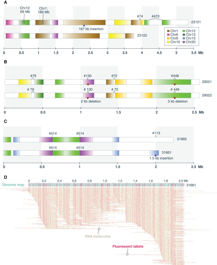
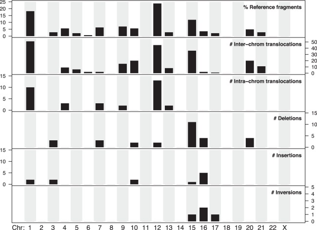
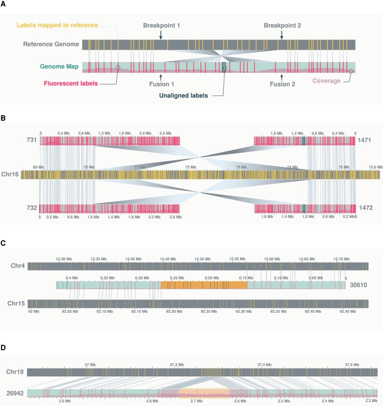
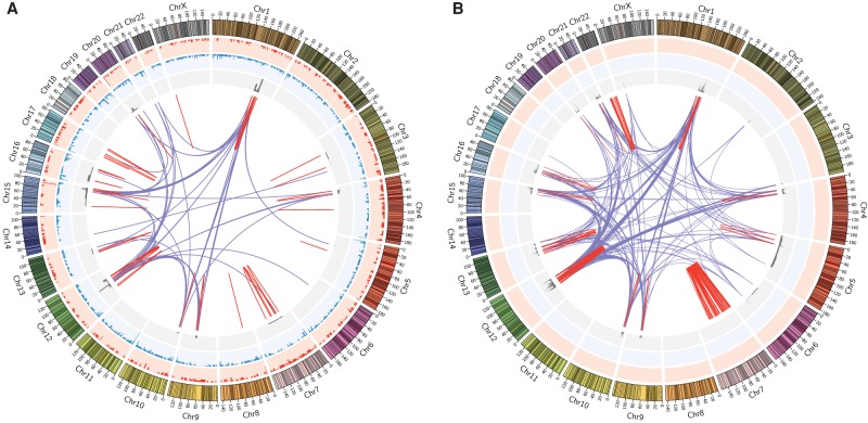
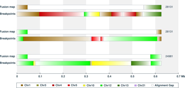
Genomic rearrangements are common in cancer, with demonstrated links to disease progression and treatment response. These rearrangements can be complex, resulting in fusions of multiple chromosomal fragments and generation of derivative chromosomes. Although methods exist for detecting individual fusions, they are generally unable to reconstruct complex chained events. To overcome these limitations, we adopted a new optical mapping approach, allowing megabase-length genome maps to be reconstructed and rearranged genomes to be visualized without loss of integrity. Whole-genome mapping (Bionano Genomics) of a well-studied highly rearranged liposarcoma cell line resulted in 3338 assembled consensus genome maps, including 72 fusion maps. These fusion maps represent 112.3 Mb of highly rearranged genomic regions, illuminating the complex architecture of chained fusions, including content, order, orientation, and size. Spanning the junction of 147 chromosomal translocations, we found a total of 28 Mb of interspersed sequences that could not be aligned to the reference genome. Traversing these interspersed sequences using short-read sequencing breakpoint calls, we were able to identify and place 399 sequencing fragments within the optical mapping gaps, thus illustrating the complementary nature of optical mapping and short-read sequencing. We demonstrate that optical mapping provides a powerful new approach for capturing a higher level of complex genomic architecture, creating a scaffold for renewed interpretation of sequencing data of particular relevance to human cancer.
基因组重排在癌症中很常见,与疾病进展和治疗反应有明确的联系。这些重排可能很复杂,导致多个染色体片段融合,并产生衍生染色体。虽然存在检测单个融合的方法,但它们通常无法重建复杂的连锁事件。为了克服这些限制,我们采用了一种新的光学作图方法,允许重建兆碱基长度的基因组图谱,并可视化重排的基因组,而不会丢失完整性。对一个经过充分研究的高度重排的脂肪肉瘤细胞系进行全基因组作图(Bionano Genomics),得到了 3338 个组装的共识基因组图谱,包括 72 个融合图谱。这些融合图谱代表了 112.3Mb 的高度重排基因组区域,阐明了连锁融合的复杂结构,包括内容、顺序、方向和大小。跨越 147 个染色体易位的交界处,我们发现了总共 28Mb 的散布序列,无法与参考基因组对齐。通过使用短读测序断点调用遍历这些散布序列,我们能够在光学作图间隙中识别和放置 399 个测序片段,从而说明了光学作图和短读测序的互补性。我们证明,光学作图提供了一种强大的新方法来捕获更高级别的复杂基因组结构,为重新解释对人类癌症特别相关的测序数据提供了支架。