Shri Bhagwan Mahavir Vitreoretinal services, 18, College Road, Sankara Nethralaya, Chennai, 600 006, Tamil Nadu, India.
Elite School of Optometry, 8 GST Road, Chennai, 600016, Tamil Nadu, India.
Sci Rep. 2018 Apr 4;8(1):5636. doi: 10.1038/s41598-018-24015-9.
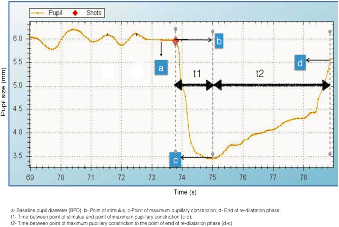
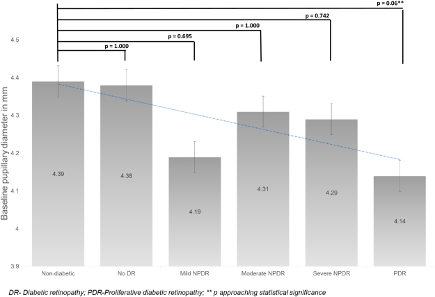
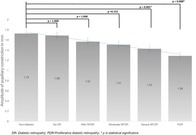
Our aim is to study the dynamics of pupillary abnormalities in varying severity of diabetic retinopathy. A non-interventional case-control study with 405 eyes of 244 subjects with diabetes, and 41 eyes of 26 subjects with no history of diabetes was done. Diabetes group was classified according to retinopathy severity: no retinopathy, mild non-proliferative diabetic retinopathy (NPDR), moderate NPDR, severe NPDR and proliferative diabetic retinopathy (PDR). After dark adaptation, pupil size and flashlight response were captured with an infrared camera. Baseline Pupil Diameter (BPD), Amplitude of Pupillary Constriction (APC), Velocity of Pupillary Constriction (VPC) and Velocity of Pupillary Dilatation (VPD). Compared to controls, mean BPD decreased with increasing severity of diabetic retinopathy. Mean APC in control group was 1.73 ± 0.37 mm and reduced in mild NPDR (1.57 ± 0.39, p = 1.000), moderate NPDR (1.51 ± 0.44, p = 0.152) and found to be significant reduced in severe NPDR (1.43 ± 0.48, p = 0.001) and PDR (1.29 ± 0.43, p = 0.008). Compared to controls, mean VPC decreased progressively with increasing severity of retinopathy, with a maximal difference in the PDR group. Mean VPD as compared to the control group was significantly reduced in the no DR (p = 0.03), mild NPDR (p = 0.038), moderate NPDR (p = 0.05), PDR group (p = 0.02). We found pupillary dynamics are abnormal in early stages of diabetic retinopathy and progress with increasing retinopathy severity.
我们的目的是研究不同严重程度糖尿病视网膜病变患者瞳孔异常的动态变化。本研究为非干预性病例对照研究,纳入了 244 名糖尿病患者的 405 只眼和 26 名无糖尿病史患者的 41 只眼。根据视网膜病变的严重程度对糖尿病组进行分类:无视网膜病变、轻度非增殖性糖尿病视网膜病变(NPDR)、中度 NPDR、重度 NPDR 和增殖性糖尿病视网膜病变(PDR)。暗适应后,用红外摄像机捕捉瞳孔大小和闪光灯反应。记录基础瞳孔直径(BPD)、瞳孔收缩幅度(APC)、瞳孔收缩速度(VPC)和瞳孔扩张速度(VPD)。与对照组相比,随着糖尿病视网膜病变严重程度的增加,平均 BPD 逐渐减小。对照组平均 APC 为 1.73±0.37mm,在轻度 NPDR 时降低(1.57±0.39,p=1.000),在中度 NPDR 时降低(1.51±0.44,p=0.152),在重度 NPDR 时显著降低(1.43±0.48,p=0.001)和 PDR 时降低(1.29±0.43,p=0.008)。与对照组相比,随着视网膜病变严重程度的增加,平均 VPC 逐渐降低,在 PDR 组差异最大。与对照组相比,无 DR 组(p=0.03)、轻度 NPDR 组(p=0.038)、中度 NPDR 组(p=0.05)、PDR 组(p=0.02)的平均 VPD 均显著降低。我们发现,糖尿病视网膜病变的早期阶段瞳孔动力学就已经异常,并随着视网膜病变严重程度的增加而进展。