• 文献检索
  • 文档翻译
  • 深度研究
  • 学术资讯
  • Suppr Zotero 插件Zotero 插件
  • 邀请有礼
  • 套餐&价格
  • 历史记录
应用&插件
Suppr Zotero 插件Zotero 插件浏览器插件Mac 客户端Windows 客户端微信小程序
定价
高级版会员购买积分包购买API积分包
服务
文献检索文档翻译深度研究API 文档MCP 服务
关于我们
关于 Suppr公司介绍联系我们用户协议隐私条款
关注我们

Suppr 超能文献

核心技术专利:CN118964589B侵权必究
粤ICP备2023148730 号-1Suppr @ 2026

文献检索

告别复杂PubMed语法,用中文像聊天一样搜索,搜遍4000万医学文献。AI智能推荐,让科研检索更轻松。

立即免费搜索

文件翻译

保留排版,准确专业,支持PDF/Word/PPT等文件格式,支持 12+语言互译。

免费翻译文档

深度研究

AI帮你快速写综述,25分钟生成高质量综述,智能提取关键信息,辅助科研写作。

立即免费体验

利用深度学习技术,在社区环境中由现场工作人员使用手持免散瞳视网膜图像检测糖尿病性视网膜病变。

Using deep learning to detect diabetic retinopathy on handheld non-mydriatic retinal images acquired by field workers in community settings.

机构信息

Institute of Ophthalmology, University College London, 11-43 Bath St., London, EC1V 9EL, UK.

Section of Ophthalmology, King's College London, London, WC2R 2LS, UK.

出版信息

Sci Rep. 2023 Jan 25;13(1):1392. doi: 10.1038/s41598-023-28347-z.

DOI:10.1038/s41598-023-28347-z
PMID:36697482
原文链接:https://pmc.ncbi.nlm.nih.gov/articles/PMC9876892/
Abstract

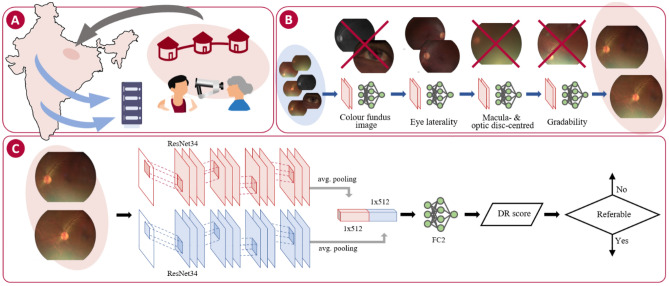
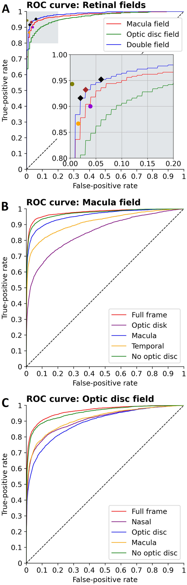
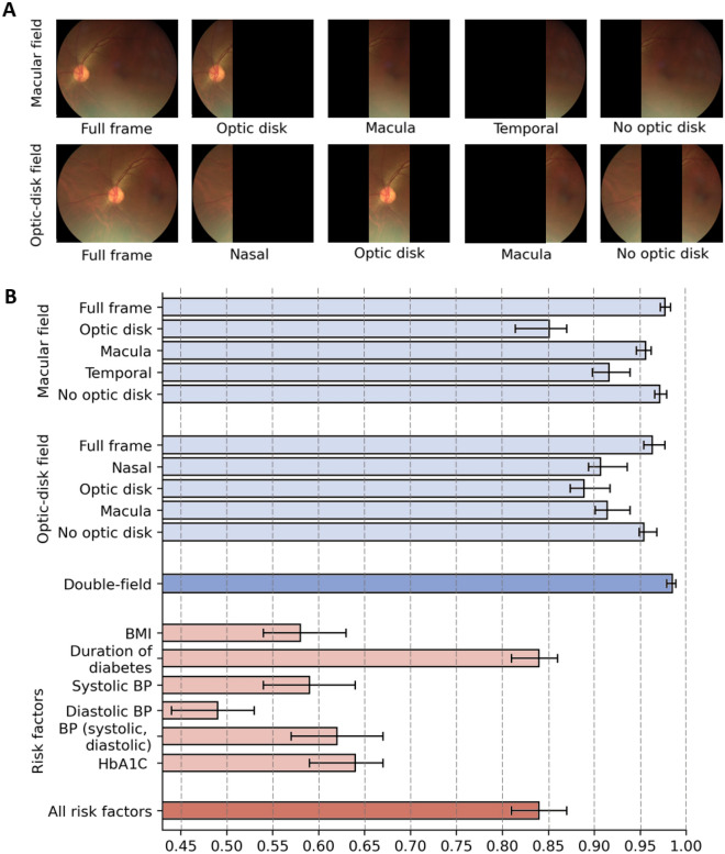
Diabetic retinopathy (DR) at risk of vision loss (referable DR) needs to be identified by retinal screening and referred to an ophthalmologist. Existing automated algorithms have mostly been developed from images acquired with high cost mydriatic retinal cameras and cannot be applied in the settings used in most low- and middle-income countries. In this prospective multicentre study, we developed a deep learning system (DLS) that detects referable DR from retinal images acquired using handheld non-mydriatic fundus camera by non-technical field workers in 20 sites across India. Macula-centred and optic-disc-centred images from 16,247 eyes (9778 participants) were used to train and cross-validate the DLS and risk factor based logistic regression models. The DLS achieved an AUROC of 0.99 (1000 times bootstrapped 95% CI 0.98-0.99) using two-field retinal images, with 93.86 (91.34-96.08) sensitivity and 96.00 (94.68-98.09) specificity at the Youden's index operational point. With single field inputs, the DLS reached AUROC of 0.98 (0.98-0.98) for the macula field and 0.96 (0.95-0.98) for the optic-disc field. Intergrader performance was 90.01 (88.95-91.01) sensitivity and 96.09 (95.72-96.42) specificity. The image based DLS outperformed all risk factor-based models. This DLS demonstrated a clinically acceptable performance for the identification of referable DR despite challenging image capture conditions.

摘要

糖尿病性视网膜病变(DR)有视力丧失风险(可转诊 DR),需要通过视网膜筛查识别,并转介给眼科医生。现有的自动化算法大多是基于高成本的散瞳视网膜相机获取的图像开发的,不能应用于大多数中低收入国家使用的环境中。在这项前瞻性多中心研究中,我们开发了一种深度学习系统(DLS),该系统可由非技术领域工作人员使用手持式非散瞳眼底相机从印度 20 个地点采集的视网膜图像中检测出可转诊 DR。使用来自 16247 只眼睛(9778 名参与者)的黄斑中心和视盘中心图像,对 DLS 和基于风险因素的逻辑回归模型进行训练和交叉验证。DLS 使用两视野视网膜图像获得了 0.99 的 AUROC(1000 次 bootstrap 95%CI 0.98-0.99),在 Youden 指数操作点的敏感性为 93.86(91.34-96.08),特异性为 96.00(94.68-98.09)。使用单视野输入,DLS 在黄斑区达到 0.98(0.98-0.98)的 AUROC,在视盘区达到 0.96(0.95-0.98)的 AUROC。分级器的性能为 90.01(88.95-91.01)的敏感性和 96.09(95.72-96.42)的特异性。基于图像的 DLS 优于所有基于风险因素的模型。尽管图像采集条件具有挑战性,但该 DLS 仍能对可转诊 DR 的识别表现出可接受的临床性能。

https://cdn.ncbi.nlm.nih.gov/pmc/blobs/92ee/9876892/ff13b6b719ca/41598_2023_28347_Fig4_HTML.jpg
https://cdn.ncbi.nlm.nih.gov/pmc/blobs/92ee/9876892/a01ff86a679a/41598_2023_28347_Fig1_HTML.jpg
https://cdn.ncbi.nlm.nih.gov/pmc/blobs/92ee/9876892/06bc60cb82f1/41598_2023_28347_Fig2_HTML.jpg
https://cdn.ncbi.nlm.nih.gov/pmc/blobs/92ee/9876892/a3c35d4d6d33/41598_2023_28347_Fig3_HTML.jpg
https://cdn.ncbi.nlm.nih.gov/pmc/blobs/92ee/9876892/ff13b6b719ca/41598_2023_28347_Fig4_HTML.jpg

相似文献

1
Using deep learning to detect diabetic retinopathy on handheld non-mydriatic retinal images acquired by field workers in community settings.利用深度学习技术,在社区环境中由现场工作人员使用手持免散瞳视网膜图像检测糖尿病性视网膜病变。
Sci Rep. 2023 Jan 25;13(1):1392. doi: 10.1038/s41598-023-28347-z.
2
Comparison of 2-Field and 5-Field Mydriatic Handheld Retinal Imaging in a Community-Based Diabetic Retinopathy Screening Program.基于社区的糖尿病视网膜病变筛查项目中 2 野和 5 野散瞳手持视网膜成像的比较。
Ophthalmologica. 2023;246(3-4):203-208. doi: 10.1159/000530903. Epub 2023 May 11.
3
Evaluating a Deep Learning Diabetic Retinopathy Grading System Developed on Mydriatic Retinal Images When Applied to Non-Mydriatic Community Screening.评估一种基于散瞳视网膜图像开发的深度学习糖尿病视网膜病变分级系统应用于非散瞳社区筛查时的情况。
J Clin Med. 2022 Jan 26;11(3):614. doi: 10.3390/jcm11030614.
4
Diagnostic test accuracy of diabetic retinopathy screening by physician graders using a hand-held non-mydriatic retinal camera at a tertiary level medical clinic.在三级医疗诊所,由医生分级人员使用手持式免散瞳视网膜相机进行糖尿病视网膜病变筛查的诊断测试准确性。
BMC Ophthalmol. 2019 Apr 8;19(1):89. doi: 10.1186/s12886-019-1092-3.
5
Deep learning for gradability classification of handheld, non-mydriatic retinal images.手持、非散瞳视网膜图像的可分级性分类的深度学习。
Sci Rep. 2021 May 4;11(1):9469. doi: 10.1038/s41598-021-89027-4.
6
One-field, two-field and five-field handheld retinal imaging compared with standard seven-field Early Treatment Diabetic Retinopathy Study photography for diabetic retinopathy screening.单视野、双视野和五视野手持视网膜成像与标准的七视野糖尿病性视网膜病变研究摄影术在糖尿病性视网膜病变筛查中的比较。
Br J Ophthalmol. 2024 May 21;108(5):735-741. doi: 10.1136/bjo-2022-321849.
7
Performance of Automated Machine Learning for Diabetic Retinopathy Image Classification from Multi-field Handheld Retinal Images.基于多视场手持视网膜图像的糖尿病性视网膜病变图像分类的自动化机器学习性能。
Ophthalmol Retina. 2023 Aug;7(8):703-712. doi: 10.1016/j.oret.2023.03.003. Epub 2023 Mar 15.
8
Comparison of Handheld Retinal Imaging with ETDRS 7-Standard Field Photography for Diabetic Retinopathy and Diabetic Macular Edema.手持式视网膜成像与 ETDRS 7 标准视野摄影在糖尿病视网膜病变和糖尿病黄斑水肿中的比较。
Ophthalmol Retina. 2022 Jul;6(7):548-556. doi: 10.1016/j.oret.2022.03.002. Epub 2022 Mar 9.
9
Sensitivity and specificity of automated analysis of single-field non-mydriatic fundus photographs by Bosch DR Algorithm-Comparison with mydriatic fundus photography (ETDRS) for screening in undiagnosed diabetic retinopathy.博世糖尿病视网膜病变(DR)算法对单视野免散瞳眼底照片进行自动分析的敏感性和特异性——与散瞳眼底摄影(ETDRS)在未诊断的糖尿病视网膜病变筛查中的比较
PLoS One. 2017 Dec 27;12(12):e0189854. doi: 10.1371/journal.pone.0189854. eCollection 2017.
10
Comparisons of handheld retinal imaging devices with ultrawide field images for determining diabetic retinopathy severity.手持式视网膜成像设备与超广角图像在确定糖尿病视网膜病变严重程度方面的比较。
Acta Ophthalmol. 2023 Sep;101(6):670-678. doi: 10.1111/aos.15651. Epub 2023 Feb 27.

引用本文的文献

1
The role of artificial intelligence in the diagnosis of diabetic retinopathy through retinal lesion features: a narrative review.通过视网膜病变特征探讨人工智能在糖尿病视网膜病变诊断中的作用:一项叙述性综述
Quant Imaging Med Surg. 2025 May 1;15(5):4816-4846. doi: 10.21037/qims-24-1791. Epub 2025 Apr 16.
2
Performance of automated machine learning in detecting fundus diseases based on ophthalmologic B-scan ultrasound images.基于眼科B超超声图像的自动机器学习在检测眼底疾病中的性能。
BMJ Open Ophthalmol. 2024 Dec 11;9(1):e001873. doi: 10.1136/bmjophth-2024-001873.
3
Novel artificial intelligence algorithms for diabetic retinopathy and diabetic macular edema.

本文引用的文献

1
Evaluating a Deep Learning Diabetic Retinopathy Grading System Developed on Mydriatic Retinal Images When Applied to Non-Mydriatic Community Screening.评估一种基于散瞳视网膜图像开发的深度学习糖尿病视网膜病变分级系统应用于非散瞳社区筛查时的情况。
J Clin Med. 2022 Jan 26;11(3):614. doi: 10.3390/jcm11030614.
2
Diabetic Retinopathy Fundus Image Classification and Lesions Localization System Using Deep Learning.基于深度学习的糖尿病视网膜病变眼底图像分类及病变定位系统
Sensors (Basel). 2021 May 26;21(11):3704. doi: 10.3390/s21113704.
3
Deep learning for gradability classification of handheld, non-mydriatic retinal images.
用于糖尿病性视网膜病变和糖尿病性黄斑水肿的新型人工智能算法。
Eye Vis (Lond). 2024 Jun 17;11(1):23. doi: 10.1186/s40662-024-00389-y.
4
Comparing code-free and bespoke deep learning approaches in ophthalmology.比较眼科领域的无代码和定制深度学习方法。
Graefes Arch Clin Exp Ophthalmol. 2024 Sep;262(9):2785-2798. doi: 10.1007/s00417-024-06432-x. Epub 2024 Mar 6.
5
Machine learning in precision diabetes care and cardiovascular risk prediction.机器学习在精准糖尿病护理和心血管风险预测中的应用。
Cardiovasc Diabetol. 2023 Sep 25;22(1):259. doi: 10.1186/s12933-023-01985-3.
6
Evaluation of a portable retinal imaging device: towards a comparative quantitative analysis for morphological measurements of retinal blood vessels.便携式视网膜成像设备的评估:迈向视网膜血管形态测量的比较定量分析
R Soc Open Sci. 2023 Jun 21;10(6):230065. doi: 10.1098/rsos.230065. eCollection 2023 Jun.
手持、非散瞳视网膜图像的可分级性分类的深度学习。
Sci Rep. 2021 May 4;11(1):9469. doi: 10.1038/s41598-021-89027-4.
4
Predicting the risk of developing diabetic retinopathy using deep learning.利用深度学习预测糖尿病视网膜病变的风险。
Lancet Digit Health. 2021 Jan;3(1):e10-e19. doi: 10.1016/S2589-7500(20)30250-8. Epub 2020 Nov 26.
5
Applications of deep learning in fundus images: A review.深度学习在眼底图像中的应用:综述。
Med Image Anal. 2021 Apr;69:101971. doi: 10.1016/j.media.2021.101971. Epub 2021 Jan 20.
6
Artificial intelligence using deep learning to screen for referable and vision-threatening diabetic retinopathy in Africa: a clinical validation study.人工智能利用深度学习在非洲筛查可转诊和威胁视力的糖尿病视网膜病变:一项临床验证研究。
Lancet Digit Health. 2019 May;1(1):e35-e44. doi: 10.1016/S2589-7500(19)30004-4. Epub 2019 May 2.
7
Protocol on a multicentre statistical and economic modelling study of risk-based stratified and personalised screening for diabetes and its complications in India (SMART India).基于风险分层和个体化的糖尿病及其并发症筛查的多中心统计和经济建模研究方案:印度(SMART India)。
BMJ Open. 2020 Dec 12;10(12):e039657. doi: 10.1136/bmjopen-2020-039657.
8
Deep Learning-Based Segmentation and Quantification of Retinal Capillary Non-Perfusion on Ultra-Wide-Field Retinal Fluorescein Angiography.基于深度学习的超广角视网膜荧光血管造影中视网膜毛细血管无灌注区的分割与量化
J Clin Med. 2020 Aug 6;9(8):2537. doi: 10.3390/jcm9082537.
9
Screening for diabetic retinopathy: new perspectives and challenges.糖尿病视网膜病变筛查:新视角与新挑战。
Lancet Diabetes Endocrinol. 2020 Apr;8(4):337-347. doi: 10.1016/S2213-8587(19)30411-5. Epub 2020 Feb 27.
10
Automatic screening of fundus images using a combination of convolutional neural network and hand-crafted features.结合卷积神经网络和手工特征自动筛查眼底图像。
Annu Int Conf IEEE Eng Med Biol Soc. 2019 Jul;2019:2699-2702. doi: 10.1109/EMBC.2019.8857073.