Ewertsen Caroline, Carlsen Jonathan, Perveez Mohammed Aftab, Schytz Henrik
Copenhagen University Hospital, Rigshospitalet, Department of Radiology, Copenhagen OE, Denmark.
Rigshospitalet, Radiologisk klinik, Kopenhagen, Denmark.
Ultrasound Int Open. 2018 Jan;4(1):E23-E29. doi: 10.1055/s-0044-102013. Epub 2018 Apr 4.
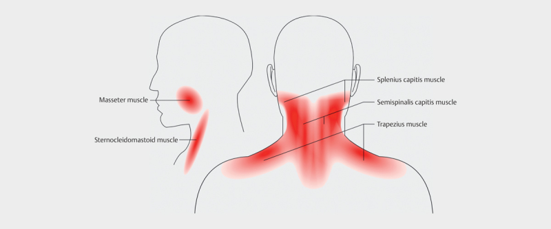
to establish reference values for ultrasound shear-wave elastography for pericranial muscles in healthy individuals (m. trapezius, m. splenius capitis, m. semispinalis capitis, m. sternocleidomastoideus and m. masseter). Also to evaluate day-to-day variations in the shear-wave speeds and evaluate the effect of the pennation of the muscle fibers, ie scanning parallel or perpendicularly to the fibers.
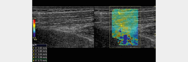
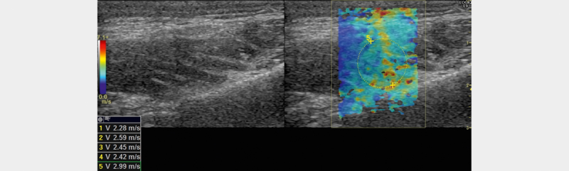
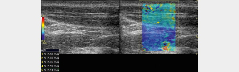
10 healthy individuals (5 males and 5 females) had their pericranial muscles examined with shear-wave elastography in two orthogonal planes on two different days for their dominant and non-dominant side. Mean shear wave speeds from 5 ROI's in each muscle, for each scan plane for the dominant and non-dominant side for the two days were calculated. The effect of the different parameters - muscle pennation, gender, dominant vs non-dominant side and day was evaluated.
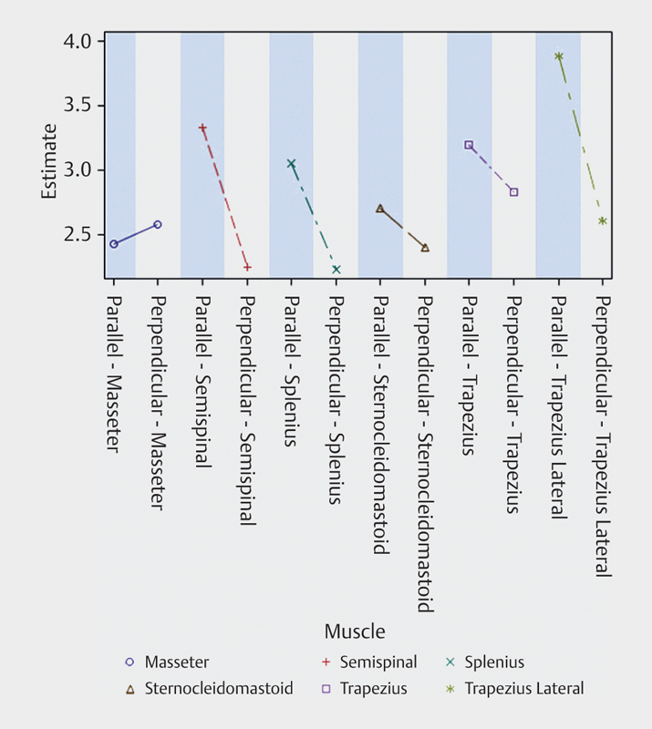
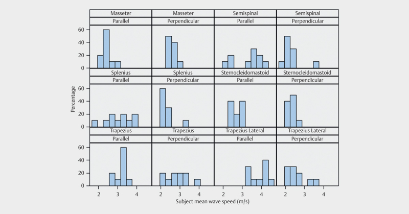
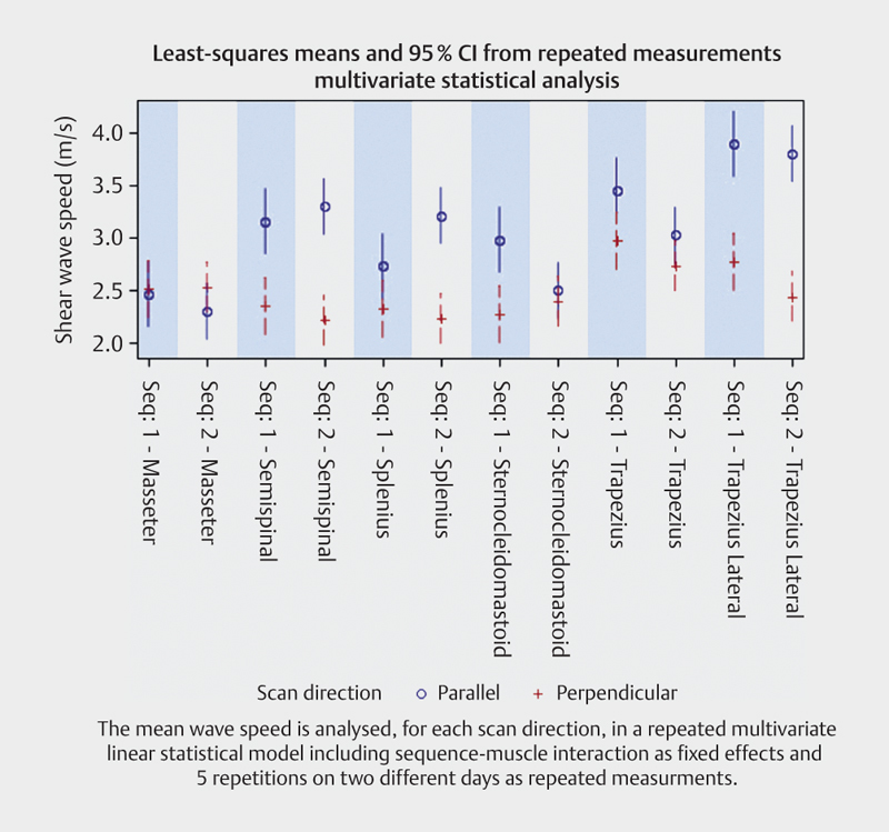
The effect of scan plane in relation to muscle pennation was statistically significant (p<0.0001). The mean shear-wave speed when scanning parallel to the muscle fibers was significantly higher than the mean shear-wave speed when scanning perpendicularly to the fibers. The day-to-day variation was statistically significant (p=0.0258), but not clinically relevant. Shear-wave speeds differed significantly between muscles. Mean shear wave speeds (m/s) for the muscles in the parallel plane were: for masseter 2.45 (SD:+/-0.25), semispinal 3.36 (SD:+/-0.75), splenius 3.04 (SD:+/-0.65), sternocleidomastoid 2.75 (SD:+/-0.23), trapezius 3.20 (SD:+/-0.27) and trapezius lateral 3.87 (SD:+/-3.87).
The shear wave speed variation depended on the direction of scanning. Shear wave elastography may be a method to evaluate muscle stiffness in patients suffering from chronic neck pain.
建立健康个体(斜方肌、头夹肌、头半棘肌、胸锁乳突肌和咬肌)颅骨周围肌肉超声剪切波弹性成像的参考值。同时评估剪切波速度的每日变化,并评估肌纤维羽状化的影响,即平行或垂直于纤维进行扫描。
10名健康个体(5名男性和5名女性)在两天内,于两个相互垂直的平面上,对其优势侧和非优势侧的颅骨周围肌肉进行剪切波弹性成像检查。计算两天内每个肌肉在每个扫描平面上,优势侧和非优势侧5个感兴趣区域(ROI)的平均剪切波速度。评估不同参数(肌肉羽状化、性别、优势侧与非优势侧以及日期)的影响。
扫描平面与肌肉羽状化的关系具有统计学意义(p<0.0001)。平行于肌纤维扫描时的平均剪切波速度显著高于垂直于纤维扫描时的平均剪切波速度。每日变化具有统计学意义(p=0.0258),但无临床相关性。不同肌肉之间的剪切波速度差异显著。平行平面上各肌肉的平均剪切波速度(m/s)分别为:咬肌2.45(标准差:±0.25),半棘肌3.36(标准差:±0.75),夹肌3.04(标准差:±0.65),胸锁乳突肌2.75(标准差:±0.23),斜方肌3.20(标准差:±0.27),斜方肌外侧3.87(标准差:±3.87)。
剪切波速度变化取决于扫描方向。剪切波弹性成像可能是评估慢性颈部疼痛患者肌肉僵硬程度的一种方法。