Cancer and Stem Cell Biology, Duke-NUS Medical School, 169857 Singapore.
Centre for Computational Biology, Duke-NUS Medical School, 169857 Singapore.
Genome Res. 2018 May;28(5):654-665. doi: 10.1101/gr.230219.117. Epub 2018 Apr 9.
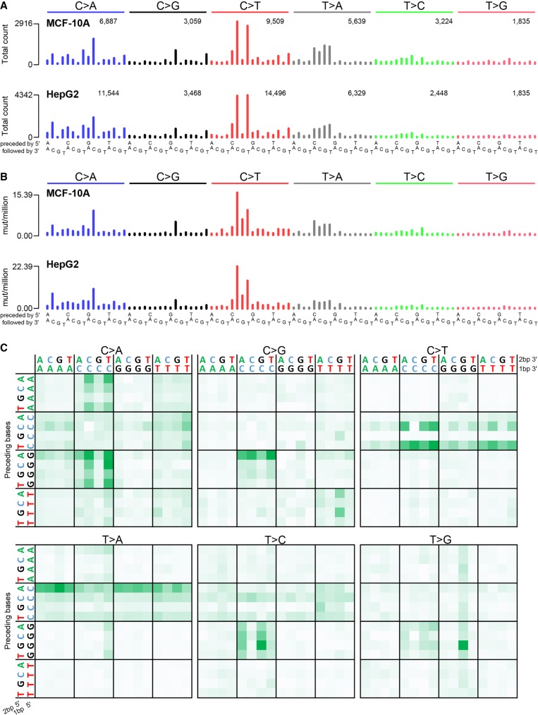
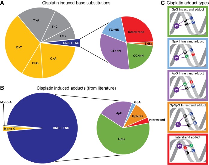
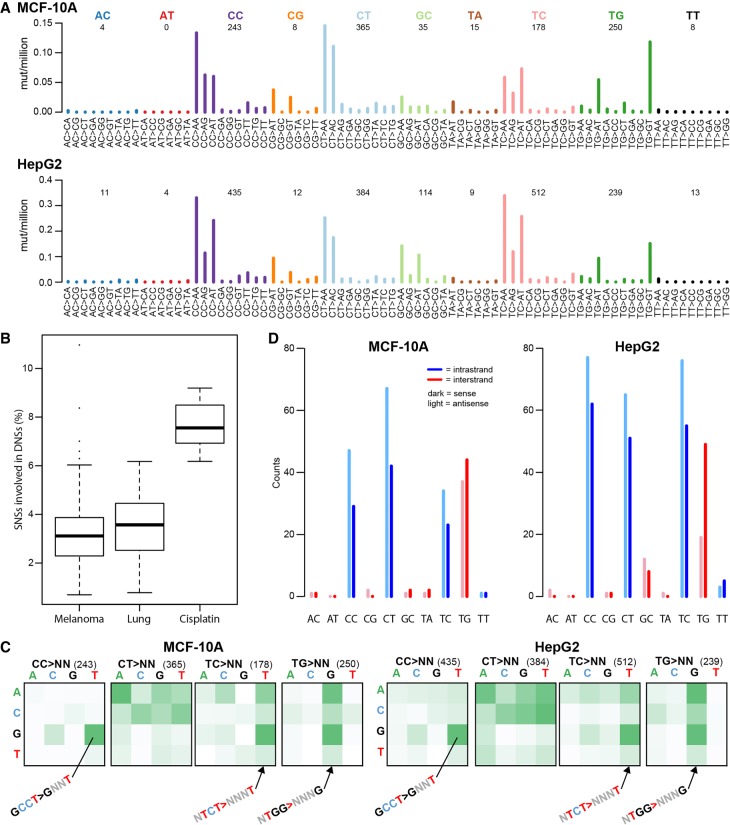
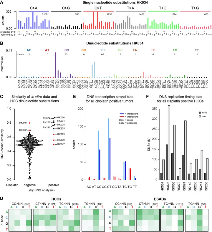
Cisplatin reacts with DNA and thereby likely generates a characteristic pattern of somatic mutations, called a mutational signature. Despite widespread use of cisplatin in cancer treatment and its role in contributing to secondary malignancies, its mutational signature has not been delineated. We hypothesize that cisplatin's mutational signature can serve as a biomarker to identify cisplatin mutagenesis in suspected secondary malignancies. Knowledge of which tissues are at risk of developing cisplatin-induced secondary malignancies could lead to guidelines for noninvasive monitoring for secondary malignancies after cisplatin chemotherapy. We performed whole genome sequencing of 10 independent clones of cisplatin-exposed MCF-10A and HepG2 cells and delineated the patterns of single and dinucleotide mutations in terms of flanking sequence, transcription strand bias, and other characteristics. We used the mSigAct signature presence test and nonnegative matrix factorization to search for cisplatin mutagenesis in hepatocellular carcinomas and esophageal adenocarcinomas. All clones showed highly consistent patterns of single and dinucleotide substitutions. The proportion of dinucleotide substitutions was high: 8.1% of single nucleotide substitutions were part of dinucleotide substitutions, presumably due to cisplatin's propensity to form intra- and interstrand crosslinks between purine bases in DNA. We identified likely cisplatin exposure in nine hepatocellular carcinomas and three esophageal adenocarcinomas. All hepatocellular carcinomas for which clinical data were available and all esophageal cancers indeed had histories of cisplatin treatment. We experimentally delineated the single and dinucleotide mutational signature of cisplatin. This signature enabled us to detect previous cisplatin exposure in human hepatocellular carcinomas and esophageal adenocarcinomas with high confidence.
顺铂与 DNA 反应,从而可能产生一种特征性的体细胞突变模式,称为突变特征。尽管顺铂在癌症治疗中广泛应用,并在导致继发性恶性肿瘤方面发挥作用,但它的突变特征尚未确定。我们假设顺铂的突变特征可以作为生物标志物,用于识别疑似继发性恶性肿瘤中的顺铂诱变。了解哪些组织有发展为顺铂诱导的继发性恶性肿瘤的风险,可能会导致在顺铂化疗后针对继发性恶性肿瘤进行非侵入性监测的指南。我们对暴露于顺铂的 MCF-10A 和 HepG2 细胞的 10 个独立克隆进行了全基因组测序,并根据侧翼序列、转录链偏向性和其他特征描绘了单核苷酸和二核苷酸突变的模式。我们使用 mSigAct 特征存在测试和非负矩阵分解在肝细胞癌和食管腺癌中搜索顺铂诱变。所有克隆均显示出高度一致的单核苷酸和二核苷酸取代模式。二核苷酸取代的比例很高:8.1%的单核苷酸取代是二核苷酸取代的一部分,这可能是由于顺铂倾向于在 DNA 中嘌呤碱基之间形成内链和链间交联。我们在 9 例肝细胞癌和 3 例食管腺癌中鉴定出可能的顺铂暴露。所有可获得临床数据的肝细胞癌和所有食管癌实际上都有顺铂治疗史。我们通过实验描绘了顺铂的单核苷酸和二核苷酸突变特征。该特征使我们能够高度确信地在人肝细胞癌和食管腺癌中检测到先前的顺铂暴露。