CICS-UBI-Health Sciences Research Centre, University of Beira Interior, 6201-506 Covilhã, Portugal.
Chemistry Department, Faculty of Sciences, University of Beira Interior, 6201-001 Covilhã, Portugal.
Int J Mol Sci. 2018 Apr 11;19(4):1157. doi: 10.3390/ijms19041157.
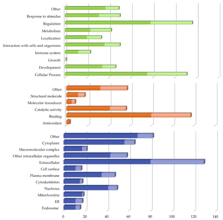
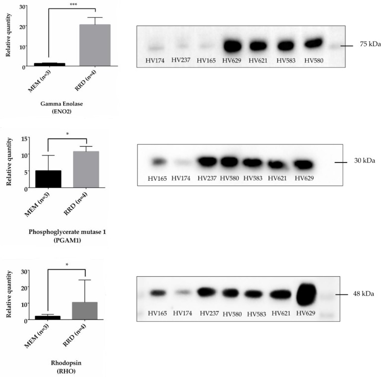
Rhegmatogenous retinal detachment (RRD) is a potentially blinding condition characterized by a physical separation between neurosensory retina and retinal pigment epithelium. Quantitative proteomics can help to understand the changes that occur at the cellular level during RRD, providing additional information about the molecular mechanisms underlying its pathogenesis. In the present study, iTRAQ labeling was combined with two-dimensional LC-ESI-MS/MS to find expression changes in the proteome of vitreous from patients with RRD when compared to control samples. A total of 150 proteins were found differentially expressed in the vitreous of patients with RRD, including 96 overexpressed and 54 underexpressed. Several overexpressed proteins, several such as glycolytic enzymes (fructose-bisphosphate aldolase A, gamma-enolase, and phosphoglycerate kinase 1), glucose transporters (GLUT-1), growth factors (metalloproteinase inhibitor 1), and serine protease inhibitors (plasminogen activator inhibitor 1) are regulated by HIF-1, which suggests that HIF-1 signaling pathway can be triggered in response to RRD. Also, the accumulation of photoreceptor proteins, including phosducin, rhodopsin, and s-arrestin, and vimentin in vitreous may indicate that photoreceptor degeneration occurs in RRD. Also, the accumulation of photoreceptor proteins, including phosducin, rhodopsin, and s-arrestin, and vimentin in vitreous may indicate that photoreceptor degeneration occurs in RRD. Nevertheless, the differentially expressed proteins found in this study suggest that different mechanisms are activated after RRD to promote the survival of retinal cells through complex cellular responses.
孔源性视网膜脱离(RRD)是一种潜在致盲的疾病,其特征是神经感觉视网膜和视网膜色素上皮之间发生物理性分离。定量蛋白质组学可以帮助理解 RRD 发生时细胞水平的变化,为其发病机制的分子机制提供更多信息。在本研究中,iTRAQ 标记与二维 LC-ESI-MS/MS 相结合,以发现与对照样品相比,RRD 患者玻璃体中蛋白质组的表达变化。在 RRD 患者的玻璃体中发现了总共 150 种差异表达的蛋白质,包括 96 种过度表达和 54 种表达不足。几种过度表达的蛋白质,如糖酵解酶(果糖二磷酸醛缩酶 A、γ-烯醇酶和磷酸甘油酸激酶 1)、葡萄糖转运蛋白(GLUT-1)、生长因子(金属蛋白酶抑制剂 1)和丝氨酸蛋白酶抑制剂(纤溶酶原激活物抑制剂 1)受 HIF-1 调节,这表明 HIF-1 信号通路可以对 RRD 做出反应。此外,视蛋白(包括视黄醛、视黄醛和 s-抑制素)和玻璃体中的波形蛋白的积累可能表明在 RRD 中发生了光感受器变性。尽管如此,本研究中发现的差异表达蛋白表明,RRD 后会激活不同的机制,通过复杂的细胞反应促进视网膜细胞的存活。