Division of Gastroenterology and Hepatology, Department of Internal Medicine III, Medical University of Vienna, Waehringer Guertel 18-20, A-1090, Vienna, Austria.
Centre for Medical Statistics, Informatics and Intelligent Systems, Medical University of Vienna, Spitalgasse 23, A-1090, Vienna, Austria.
Sci Rep. 2018 Apr 18;8(1):6220. doi: 10.1038/s41598-018-24437-5.
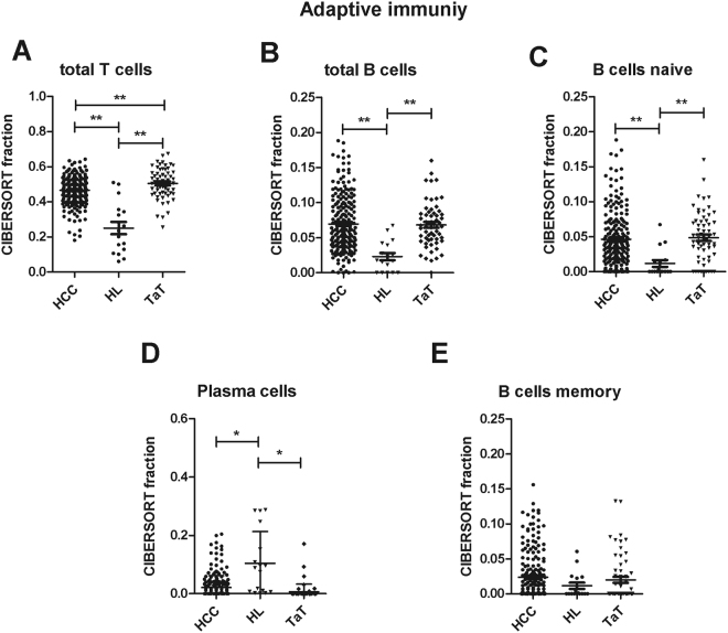
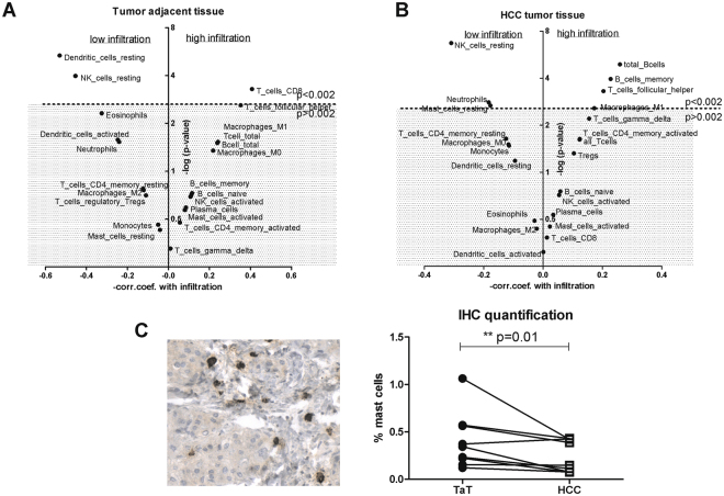
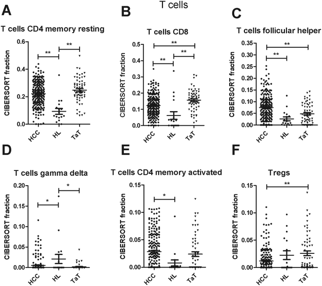
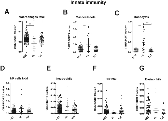
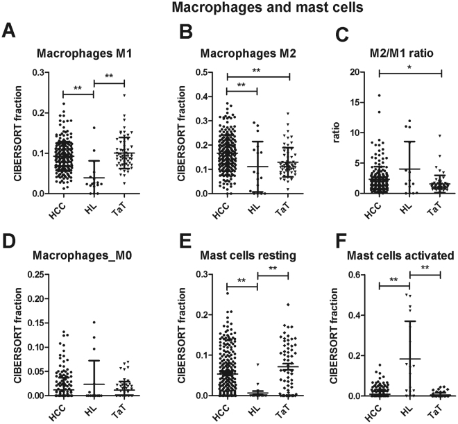
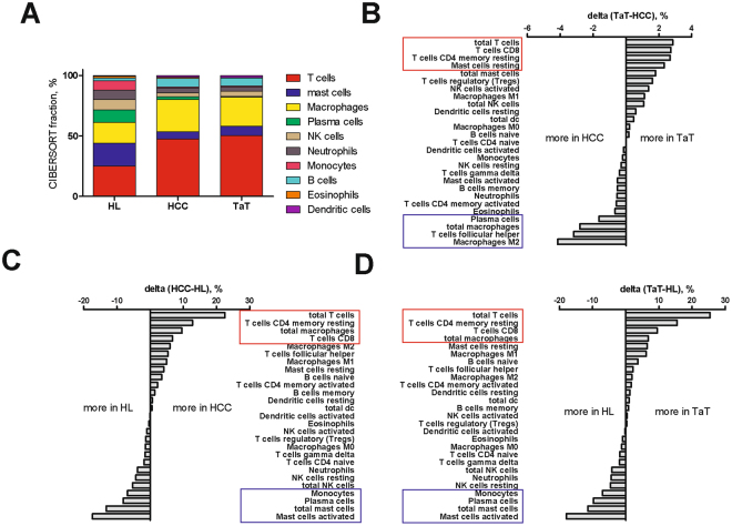
Tumor-infiltrating immune cells are highly relevant for prognosis and identification of immunotherapy targets in hepatocellular carcinoma (HCC). The recently developed CIBERSORT method allows immune cell profiling by deconvolution of gene expression microarray data. By applying CIBERSORT, we assessed the relative proportions of immune cells in 41 healthy human livers, 305 HCC samples and 82 HCC adjacent tissues. The obtained immune cell profiles provided enumeration and activation status of 22 immune cell subtypes. Mast cells were evaluated by immunohistochemistry in ten HCC patients. Activated mast cells, monocytes and plasma cells were decreased in HCC, while resting mast cells, total and naïve B cells, CD4 memory resting and CD8 T cells were increased when compared to healthy livers. Previously described S1, S2 and S3 molecular HCC subclasses demonstrated increased M1-polarized macrophages in the S3 subclass with good prognosis. Strong total immune cell infiltration into HCC correlated with total B cells, memory B cells, T follicular helper cells and M1 macrophages, whereas weak infiltration was linked to resting NK cells, neutrophils and resting mast cells. Immunohistochemical analysis of patient samples confirmed the reduced frequency of mast cells in human HCC tumor tissue as compared to tumor adjacent tissue. Our data demonstrate that deconvolution of gene expression data by CIBERSORT provides valuable information about immune cell composition of HCC patients.
肿瘤浸润免疫细胞与肝癌(HCC)的预后和免疫治疗靶点的鉴定密切相关。最近开发的 CIBERSORT 方法允许通过基因表达微阵列数据的反卷积来进行免疫细胞分析。通过应用 CIBERSORT,我们评估了 41 个健康人肝、305 个 HCC 样本和 82 个 HCC 相邻组织中免疫细胞的相对比例。获得的免疫细胞图谱提供了 22 种免疫细胞亚型的计数和激活状态。在 10 名 HCC 患者中通过免疫组织化学评估肥大细胞。与健康肝脏相比,肥大细胞、单核细胞和浆细胞在 HCC 中减少,而静止肥大细胞、总和幼稚 B 细胞、CD4 记忆静止和 CD8 T 细胞增加。先前描述的 S1、S2 和 S3 分子 HCC 亚类在具有良好预后的 S3 亚类中显示出 M1 极化巨噬细胞增加。HCC 中的总免疫细胞浸润与总 B 细胞、记忆 B 细胞、滤泡辅助 T 细胞和 M1 巨噬细胞有关,而弱浸润与静止 NK 细胞、中性粒细胞和静止肥大细胞有关。对患者样本的免疫组织化学分析证实,与肿瘤相邻组织相比,肥大细胞在人 HCC 肿瘤组织中的频率降低。我们的数据表明,CIBERSORT 通过基因表达数据的反卷积提供了有关 HCC 患者免疫细胞组成的有价值信息。