Shapiro L M, Gibson D G
National Heart Hospital, London.
Br Heart J. 1988 Apr;59(4):438-45. doi: 10.1136/hrt.59.4.438.
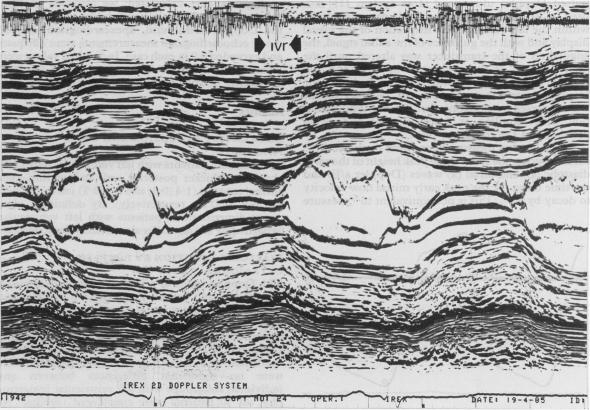
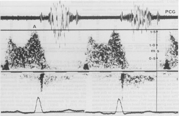
The relative sensitivities of and interrelations between different measurements of diastolic function were studied in 50 patients with left ventricular hypertrophy diagnosed on anatomical grounds. Isovolumic relaxation time, the interval from minimum cavity dimension to mitral valve opening and relative dimension increase during this period, and the peak rate of dimension increase and wall thinning during rapid ventricular filling were measured by digitised M mode echocardiography. The relative heights of peak early diastolic and atrial velocities (a/E) and the time for decline of early diastolic velocity to half its peak value (velocity half time) were measured on continuous wave and pulsed Doppler and the relative height of the "a" wave was measured by apexcardiogram. All sets of values except those of the interval from minimum dimension to mitral opening were unimodally distributed, and all differed significantly from those in 20 age matched controls. The relative height of the "a" wave on the apexcardiogram (90% values were abnormal) was the most sensitive method of studying left ventricular diastolic function and peak rate of dimension increase was the least sensitive. Though none of the correlations was high, there were individual associations between peak rate of dimension increase, a/E, peak wall thinning rate, and velocity half time, and independently between delay in mitral valve opening and dimension change during this period. Other values seemed to be independent of one another, suggesting a different physiological basis. It is concluded that these various abnormal values do not reflect a single underlying disturbance of diastolic function. There are at least four possible discrete abnormalities: prolongation of isovolumic relaxation; incoordination during isovolumic relaxation; reduced rate of rapid filling; and an increase in the relative amplitude of the "a" wave probably caused by increased passive stiffness. These may be present singly or in combination in any patient.
对50例经解剖学诊断为左心室肥厚的患者,研究了不同舒张功能测量方法之间的相对敏感性及相互关系。采用数字化M型超声心动图测量等容舒张时间、从最小腔径至二尖瓣开放的间期以及此期间的相对径增加量,还有快速心室充盈期的径增加峰值速率和室壁变薄速率。通过连续波和脉冲多普勒测量舒张早期峰值和心房速度的相对高度(a/E)以及舒张早期速度降至其峰值一半所需的时间(速度减半时间),通过心尖搏动图测量“a”波的相对高度。除了从最小径至二尖瓣开放的间期外,所有数值组均呈单峰分布,且与20例年龄匹配的对照组均有显著差异。心尖搏动图上“a”波的相对高度(90%的值异常)是研究左心室舒张功能最敏感的方法,径增加峰值速率是最不敏感的。虽然各相关性均不高,但径增加峰值速率、a/E、室壁变薄峰值速率和速度减半时间之间存在个体关联,二尖瓣开放延迟与此期间的径变化之间也独立存在关联。其他数值似乎相互独立,提示存在不同的生理基础。结论是,这些各种异常值并不反映舒张功能的单一潜在紊乱。至少有四种可能的离散异常:等容舒张期延长;等容舒张期不协调;快速充盈速率降低;以及“a”波相对振幅增加,可能是由被动僵硬度增加所致。这些异常可能单独或联合出现在任何患者中。