• 文献检索
  • 文档翻译
  • 深度研究
  • 学术资讯
  • Suppr Zotero 插件Zotero 插件
  • 邀请有礼
  • 套餐&价格
  • 历史记录
应用&插件
Suppr Zotero 插件Zotero 插件浏览器插件Mac 客户端Windows 客户端微信小程序
定价
高级版会员购买积分包购买API积分包
服务
文献检索文档翻译深度研究API 文档MCP 服务
关于我们
关于 Suppr公司介绍联系我们用户协议隐私条款
关注我们

Suppr 超能文献

核心技术专利:CN118964589B侵权必究
粤ICP备2023148730 号-1Suppr @ 2026

文献检索

告别复杂PubMed语法,用中文像聊天一样搜索,搜遍4000万医学文献。AI智能推荐,让科研检索更轻松。

立即免费搜索

文件翻译

保留排版,准确专业,支持PDF/Word/PPT等文件格式,支持 12+语言互译。

免费翻译文档

深度研究

AI帮你快速写综述,25分钟生成高质量综述,智能提取关键信息,辅助科研写作。

立即免费体验

玻璃体切片的相关显微镜检查为 BAR 结构域的原位组织提供了深入了解。

Correlative Microscopy of Vitreous Sections Provides Insights into BAR-Domain Organization In Situ.

机构信息

Sir William Dunn School of Pathology, University of Oxford, South Parks Road, Oxford OX1 3RE, UK; Central Oxford Structural and Molecular Imaging Centre, South Parks Road, Oxford OX1 3RE, UK; Structural Studies Division, MRC Laboratory of Molecular Biology, Francis Crick Avenue, Cambridge CB2 0QH, UK.

Cell Biology Division, MRC Laboratory of Molecular Biology, Francis Crick Avenue, Cambridge CB2 0QH, UK.

出版信息

Structure. 2018 Jun 5;26(6):879-886.e3. doi: 10.1016/j.str.2018.03.015. Epub 2018 Apr 19.

DOI:10.1016/j.str.2018.03.015
PMID:29681471
原文链接:https://pmc.ncbi.nlm.nih.gov/articles/PMC5992340/
Abstract

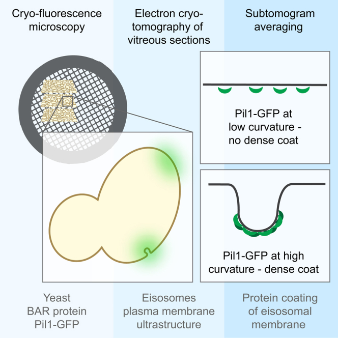
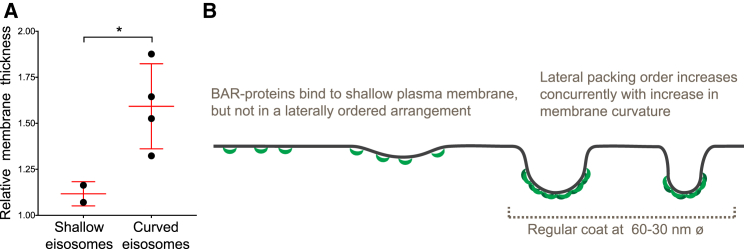
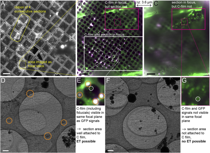
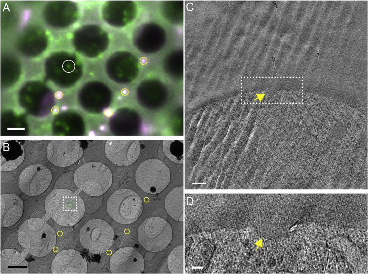
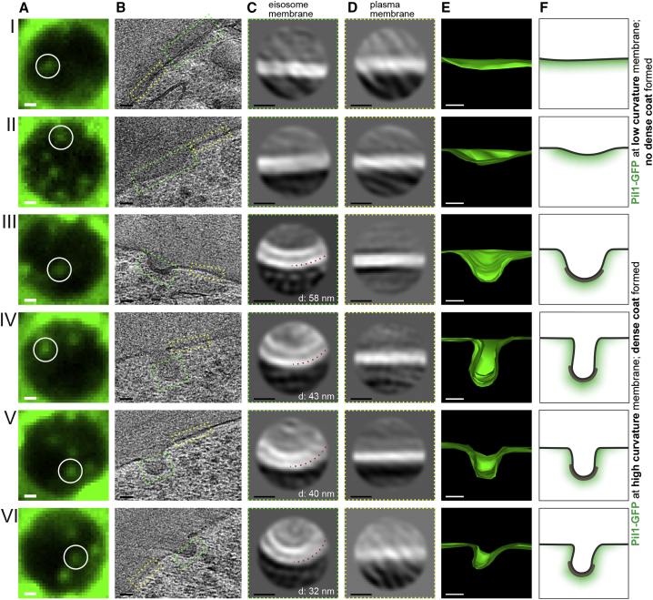
Electron microscopy imaging of macromolecular complexes in their native cellular context is limited by the inherent difficulty to acquire high-resolution tomographic data from thick cells and to specifically identify elusive structures within crowded cellular environments. Here, we combined cryo-fluorescence microscopy with electron cryo-tomography of vitreous sections into a coherent correlative microscopy workflow, ideal for detection and structural analysis of elusive protein assemblies in situ. We used this workflow to address an open question on BAR-domain coating of yeast plasma membrane compartments known as eisosomes. BAR domains can sense or induce membrane curvature, and form scaffold-like membrane coats in vitro. Our results demonstrate that in cells, the BAR protein Pil1 localizes to eisosomes of varying membrane curvature. Sub-tomogram analysis revealed a dense protein coat on curved eisosomes, which was not present on shallow eisosomes, indicating that while BAR domains can assemble at shallow membranes in vivo, scaffold formation is tightly coupled to curvature generation.

摘要

在天然细胞环境中对大分子复合物进行电子显微镜成像受到固有困难的限制,难以从厚细胞中获取高分辨率的断层扫描数据,并且难以在拥挤的细胞环境中特异性识别难以捉摸的结构。在这里,我们将冷冻荧光显微镜与玻璃切片的电子冷冻断层扫描结合到一个连贯的相关显微镜工作流程中,非常适合用于原位检测和分析难以捉摸的蛋白质组装体。我们使用该工作流程解决了关于酵母质膜隔室(称为芽殖后体)的 BAR 域涂层的一个悬而未决的问题。BAR 结构域可以感知或诱导膜曲率,并在体外形成支架样膜涂层。我们的结果表明,在细胞中,BAR 蛋白 Pil1 定位于具有不同膜曲率的芽殖后体上。子断层分析显示,在弯曲的芽殖后体上存在密集的蛋白质涂层,而在浅的芽殖后体上不存在,这表明尽管 BAR 结构域可以在体内组装在浅膜上,但支架形成与曲率生成紧密相关。

https://cdn.ncbi.nlm.nih.gov/pmc/blobs/4507/5992340/6fcb7b650ad9/gr4.jpg
https://cdn.ncbi.nlm.nih.gov/pmc/blobs/4507/5992340/e5e33d59d929/fx1.jpg
https://cdn.ncbi.nlm.nih.gov/pmc/blobs/4507/5992340/b00f657d5afd/gr1.jpg
https://cdn.ncbi.nlm.nih.gov/pmc/blobs/4507/5992340/09e484a5b93b/gr2.jpg
https://cdn.ncbi.nlm.nih.gov/pmc/blobs/4507/5992340/28e5f2f98b63/gr3.jpg
https://cdn.ncbi.nlm.nih.gov/pmc/blobs/4507/5992340/6fcb7b650ad9/gr4.jpg

相似文献

1
Correlative Microscopy of Vitreous Sections Provides Insights into BAR-Domain Organization In Situ.玻璃体切片的相关显微镜检查为 BAR 结构域的原位组织提供了深入了解。
Structure. 2018 Jun 5;26(6):879-886.e3. doi: 10.1016/j.str.2018.03.015. Epub 2018 Apr 19.
2
Eisosome proteins assemble into a membrane scaffold.质膜体蛋白装配成一个膜支架。
J Cell Biol. 2011 Nov 28;195(5):889-902. doi: 10.1083/jcb.201104040.
3
Eisosome-driven plasma membrane organization is mediated by BAR domains.Eisosome 驱动的质膜组织由 BAR 结构域介导。
Nat Struct Mol Biol. 2011 Jun 19;18(7):854-6. doi: 10.1038/nsmb.2080.
4
Cryo-EM architecture of a near-native stretch-sensitive membrane microdomain.近天然拉伸敏感膜微区的冷冻电镜结构。
Nature. 2024 Aug;632(8025):664-671. doi: 10.1038/s41586-024-07720-6. Epub 2024 Jul 24.
5
The eisosome core is composed of BAR domain proteins.质膜内板核心由 BAR 结构域蛋白组成。
Mol Biol Cell. 2011 Jul 1;22(13):2360-72. doi: 10.1091/mbc.E10-12-1021. Epub 2011 May 18.
6
Eisosomes are dynamic plasma membrane domains showing pil1-lsp1 heteroligomer binding equilibrium.埃兹体是显示pil1-lsp1异源寡聚体结合平衡的动态质膜结构域。
Biophys J. 2015 Apr 7;108(7):1633-1644. doi: 10.1016/j.bpj.2015.02.011.
7
Lsp1 partially substitutes for Pil1 function in eisosome assembly under stress conditions.Lsp1 在应激条件下部分替代 Pil1 在 eisosome 组装中的功能。
J Cell Sci. 2023 Feb 1;136(3). doi: 10.1242/jcs.260554. Epub 2023 Feb 6.
8
Single-molecule imaging of the BAR-domain protein Pil1p reveals filament-end dynamics.BAR结构域蛋白Pil1p的单分子成像揭示了细丝末端动力学。
Mol Biol Cell. 2017 Aug 15;28(17):2251-2259. doi: 10.1091/mbc.E17-04-0238. Epub 2017 Jun 28.
9
Eisosomes mark static sites of endocytosis.内质体标记内吞作用的静止位点。
Nature. 2006 Feb 23;439(7079):998-1003. doi: 10.1038/nature04472.
10
Pil1 controls eisosome biogenesis.Pil1控制内质体的生物发生。
Mol Biol Cell. 2009 Feb;20(3):809-18. doi: 10.1091/mbc.e08-03-0313. Epub 2008 Nov 26.

引用本文的文献

1
In situ high-resolution cryo-EM reconstructions from CEMOVIS.来自CEMOVIS的原位高分辨率冷冻电镜重建。
IUCrJ. 2025 Jul 1;12(Pt 4):502-510. doi: 10.1107/S2052252525005196.
2
Recovery of plasma membrane tension after a hyperosmotic shock.高渗休克后质膜张力的恢复。
Mol Biol Cell. 2025 Apr 1;36(4):ar45. doi: 10.1091/mbc.E24-10-0436. Epub 2025 Feb 19.
3
Structural biology inside multicellular specimens using electron cryotomography.利用电子冷冻断层扫描技术研究多细胞标本内部的结构生物学。

本文引用的文献

1
Single-molecule imaging of the BAR-domain protein Pil1p reveals filament-end dynamics.BAR结构域蛋白Pil1p的单分子成像揭示了细丝末端动力学。
Mol Biol Cell. 2017 Aug 15;28(17):2251-2259. doi: 10.1091/mbc.E17-04-0238. Epub 2017 Jun 28.
2
triCLEM: Combining high-precision, room temperature CLEM with cryo-fluorescence microscopy to identify very rare events.triCLEM:将高精度室温细胞光镜电镜关联技术与低温荧光显微镜相结合以识别极其罕见的事件。
Methods Cell Biol. 2017;140:303-320. doi: 10.1016/bs.mcb.2017.03.009. Epub 2017 Apr 18.
3
Correlated fluorescence microscopy and cryo-electron tomography of virus-infected or transfected mammalian cells.
Q Rev Biophys. 2025 Jan 13;58:e6. doi: 10.1017/S0033583525000010.
4
Definition of phosphatidylinositol 4,5-bisphosphate distribution by freeze-fracture replica labeling.通过冷冻断裂复型标记定义磷脂酰肌醇 4,5-二磷酸的分布。
J Cell Biol. 2025 Jan 6;224(1). doi: 10.1083/jcb.202311067. Epub 2024 Nov 4.
5
Cryo-EM architecture of a near-native stretch-sensitive membrane microdomain.近天然拉伸敏感膜微区的冷冻电镜结构。
Nature. 2024 Aug;632(8025):664-671. doi: 10.1038/s41586-024-07720-6. Epub 2024 Jul 24.
6
Lift-out cryo-FIBSEM and cryo-ET reveal the ultrastructural landscape of extracellular matrix.提取冷冻-FIBSEM 和冷冻-ET 揭示细胞外基质的超微结构景观。
J Cell Biol. 2024 Jun 3;223(6). doi: 10.1083/jcb.202309125. Epub 2024 Mar 20.
7
In situ architecture of the ER-mitochondria encounter structure.内质网-线粒体接触结构的原位结构。
Nature. 2023 Jun;618(7963):188-192. doi: 10.1038/s41586-023-06050-3. Epub 2023 May 10.
8
High-resolution structure determination using high-throughput electron cryo-tomography.利用高通量电子冷冻断层成像技术进行高分辨率结构测定。
Acta Crystallogr D Struct Biol. 2022 Jul 1;78(Pt 7):817-824. doi: 10.1107/S2059798322005010. Epub 2022 Jun 7.
9
Better, Faster, Cheaper: Recent Advances in Cryo-Electron Microscopy.更好、更快、更便宜:冷冻电镜的最新进展。
Annu Rev Biochem. 2022 Jun 21;91:1-32. doi: 10.1146/annurev-biochem-032620-110705. Epub 2022 Mar 23.
10
Preservation of Fluorescence Signal and Imaging Optimization for Integrated Light and Electron Microscopy.用于集成光镜和电镜的荧光信号保存与成像优化
Front Cell Dev Biol. 2021 Dec 15;9:737621. doi: 10.3389/fcell.2021.737621. eCollection 2021.
病毒感染或转染的哺乳动物细胞的相关荧光显微镜和 cryo-electron 断层扫描。
Nat Protoc. 2017 Jan;12(1):150-167. doi: 10.1038/nprot.2016.168. Epub 2016 Dec 15.
4
Resolving macromolecular structures from electron cryo-tomography data using subtomogram averaging in RELION.使用 RELION 中的子断层平均法从电子冷冻断层扫描数据中解析大分子结构。
Nat Protoc. 2016 Nov;11(11):2054-65. doi: 10.1038/nprot.2016.124. Epub 2016 Sep 29.
5
Cryo-Electron Tomography: Can it Reveal the Molecular Sociology of Cells in Atomic Detail?冷冻电子断层扫描:能否以原子级细节揭示细胞的分子社会学?
Trends Cell Biol. 2016 Nov;26(11):825-837. doi: 10.1016/j.tcb.2016.08.006. Epub 2016 Sep 23.
6
New hardware and workflows for semi-automated correlative cryo-fluorescence and cryo-electron microscopy/tomography.用于半自动相关低温荧光和低温电子显微镜/断层扫描的新硬件和工作流程。
J Struct Biol. 2017 Feb;197(2):83-93. doi: 10.1016/j.jsb.2016.06.020. Epub 2016 Jun 28.
7
Implementation of a cryo-electron tomography tilt-scheme optimized for high resolution subtomogram averaging.一种针对高分辨率亚断层平均优化的冷冻电子断层扫描倾斜方案的实施。
J Struct Biol. 2017 Feb;197(2):191-198. doi: 10.1016/j.jsb.2016.06.007. Epub 2016 Jun 14.
8
EMPIAR: a public archive for raw electron microscopy image data.EMPIAR:原始电子显微镜图像数据的公共存档库。
Nat Methods. 2016 May;13(5):387-8. doi: 10.1038/nmeth.3806. Epub 2016 Mar 21.
9
Supramolecular organization of the human N-BAR domain in shaping the sarcolemma membrane.人类N-BAR结构域在塑造肌膜中的超分子组织。
J Struct Biol. 2016 Jun;194(3):375-82. doi: 10.1016/j.jsb.2016.03.017. Epub 2016 Mar 22.
10
Site-Specific Cryo-focused Ion Beam Sample Preparation Guided by 3D Correlative Microscopy.由三维相关显微镜引导的位点特异性冷冻聚焦离子束样品制备
Biophys J. 2016 Feb 23;110(4):860-9. doi: 10.1016/j.bpj.2015.10.053. Epub 2016 Jan 5.