• 文献检索
  • 文档翻译
  • 深度研究
  • 学术资讯
  • Suppr Zotero 插件Zotero 插件
  • 邀请有礼
  • 套餐&价格
  • 历史记录
应用&插件
Suppr Zotero 插件Zotero 插件浏览器插件Mac 客户端Windows 客户端微信小程序
定价
高级版会员购买积分包购买API积分包
服务
文献检索文档翻译深度研究API 文档MCP 服务
关于我们
关于 Suppr公司介绍联系我们用户协议隐私条款
关注我们

Suppr 超能文献

核心技术专利:CN118964589B侵权必究
粤ICP备2023148730 号-1Suppr @ 2026

文献检索

告别复杂PubMed语法,用中文像聊天一样搜索,搜遍4000万医学文献。AI智能推荐,让科研检索更轻松。

立即免费搜索

文件翻译

保留排版,准确专业,支持PDF/Word/PPT等文件格式,支持 12+语言互译。

免费翻译文档

深度研究

AI帮你快速写综述,25分钟生成高质量综述,智能提取关键信息,辅助科研写作。

立即免费体验

单侧搏动性耳鸣早期患者的神经解剖结构改变:基于体素的形态测量学研究。

Neuroanatomical Alterations in Patients with Early Stage of Unilateral Pulsatile Tinnitus: A Voxel-Based Morphometry Study.

机构信息

Institute for Biomedical Engineering and College of Information Engineering, China Jiliang University, Hangzhou, China.

Department of Radiology, Beijing Friendship Hospital, Capital Medical University, Beijing, China.

出版信息

Neural Plast. 2018 Feb 28;2018:4756471. doi: 10.1155/2018/4756471. eCollection 2018.

DOI:10.1155/2018/4756471
PMID:29681925
原文链接:https://pmc.ncbi.nlm.nih.gov/articles/PMC5851320/
Abstract

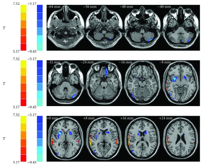
During the past several years, the rapid development of neuroimaging techniques has contributed greatly in the noninvasive imaging studies of tinnitus. The aim of the present study was to explore the brain anatomical alterations in patients with right-sided unilateral pulsatile tinnitus (PT) in the early stage of PT symptom using voxel-based morphometry (VBM) analysis. Twenty-four patients with right-sided pulsatile tinnitus and 24 age- and gender-matched normal controls were recruited to this study. Structural image data preprocessing was performed using VBM8 toolbox. Tinnitus Handicap Inventory (THI) score was acquired in the tinnitus group to assess the severity of tinnitus and tinnitus-related distress. Two-sample -test and Pearson's correlation analysis were used in statistical analysis. Patients with unilateral pulsatile tinnitus had significantly increased gray matter (GM) volume in bilateral superior temporal gyrus compared with the normal controls. However, the left cerebellum posterior lobe, left frontal superior orbital lobe (gyrus rectus), right middle occipital gyrus (MOG), and bilateral putamen showed significantly decreased brain volumes. This was the first study which demonstrated the features of neuroanatomical changes in patients with unilateral PT during their early stages of the symptom.

摘要

在过去的几年中,神经影像学技术的快速发展极大地促进了非侵入性耳鸣影像学研究。本研究旨在使用基于体素的形态测量学(VBM)分析探讨早期右侧搏动性耳鸣(PT)患者的脑解剖结构改变。本研究纳入了 24 例右侧搏动性耳鸣患者和 24 名年龄和性别匹配的正常对照者。使用 VBM8 工具箱对结构图像数据进行预处理。在耳鸣组中获取耳鸣残疾量表(THI)评分,以评估耳鸣的严重程度和与耳鸣相关的困扰。在统计分析中使用了双样本 t 检验和 Pearson 相关分析。与正常对照组相比,单侧搏动性耳鸣患者双侧颞上回的灰质(GM)体积明显增加。然而,左侧小脑后叶、左侧额上直回、右侧中枕回(MOG)和双侧壳核的脑体积明显减少。这是第一项研究,表明单侧 PT 患者在症状早期的神经解剖结构变化特征。

https://cdn.ncbi.nlm.nih.gov/pmc/blobs/446f/5851320/b3104296418e/NP2018-4756471.001.jpg

相似文献

1
Neuroanatomical Alterations in Patients with Early Stage of Unilateral Pulsatile Tinnitus: A Voxel-Based Morphometry Study.单侧搏动性耳鸣早期患者的神经解剖结构改变:基于体素的形态测量学研究。
Neural Plast. 2018 Feb 28;2018:4756471. doi: 10.1155/2018/4756471. eCollection 2018.
2
Disrupted neural activity in unilateral vascular pulsatile tinnitus patients in the early stage of disease: evidence from resting-state fMRI.单侧血管搏动性耳鸣患者疾病早期的神经活动紊乱:静息态功能磁共振成像证据
Prog Neuropsychopharmacol Biol Psychiatry. 2015 Jun 3;59:91-99. doi: 10.1016/j.pnpbp.2015.01.013. Epub 2015 Jan 30.
3
Long-term reactions to pulsatile tinnitus are marked by weakened short-range functional connectivity within a brain network in the right temporal lobe.长期的脉冲性耳鸣反应表现为右颞叶脑网络内短程功能连接减弱。
J Magn Reson Imaging. 2019 Jun;49(6):1629-1637. doi: 10.1002/jmri.26545. Epub 2018 Dec 21.
4
Abnormal resting-state functional connectivity study in unilateral pulsatile tinnitus patients with single etiology: A seed-based functional connectivity study.单病因单侧搏动性耳鸣患者静息态功能连接异常研究:基于种子点的功能连接研究。
Eur J Radiol. 2016 Nov;85(11):2023-2029. doi: 10.1016/j.ejrad.2016.09.011. Epub 2016 Sep 13.
5
Resting-state functional connectivity density mapping of etiology confirmed unilateral pulsatile tinnitus patients: Altered functional hubs in the early stage of disease.病因确诊的单侧搏动性耳鸣患者静息态功能连接密度图谱:疾病早期功能枢纽的改变
Neuroscience. 2015 Dec 3;310:27-37. doi: 10.1016/j.neuroscience.2015.09.032. Epub 2015 Sep 16.
6
Neuroanatomical Alterations in Patients With Tinnitus Before and After Sound Therapy: A Voxel-Based Morphometry Study.耳鸣患者声音治疗前后的神经解剖学改变:基于体素的形态学研究
Front Neurosci. 2020 Sep 8;14:911. doi: 10.3389/fnins.2020.00911. eCollection 2020.
7
Neuroanatomical Alterations in Patients With Tinnitus Before and After Sound Therapy: A Combined VBM and SCN Study.耳鸣患者声音治疗前后的神经解剖学改变:一项基于体素形态学和静息态脑网络的联合研究
Front Hum Neurosci. 2021 Jan 18;14:607452. doi: 10.3389/fnhum.2020.607452. eCollection 2020.
8
Outcomes at 6 months are related to brain structural and white matter microstructural reorganization in idiopathic tinnitus patients treated with sound therapy.耳鸣患者接受声音治疗后 6 个月的疗效与大脑结构和白质微观结构重组有关。
Hum Brain Mapp. 2021 Feb 15;42(3):753-765. doi: 10.1002/hbm.25260. Epub 2020 Oct 24.
9
Abnormal regional activity and functional connectivity in resting-state brain networks associated with etiology confirmed unilateral pulsatile tinnitus in the early stage of disease.与病因相关的静息态脑网络中异常的区域活动和功能连接在疾病早期证实了单侧搏动性耳鸣。
Hear Res. 2017 Mar;346:55-61. doi: 10.1016/j.heares.2017.02.004. Epub 2017 Feb 7.
10
Impairments of cingulated cortex in the generalized tonic-clonic seizure epilepsy by combining morphological and functional connectivity magnetic resonance imaging.通过结合形态学和功能连接磁共振成像研究全身强直阵挛性癫痫中扣带回皮质的损伤情况。
J Integr Neurosci. 2017;16(4):429-439. doi: 10.3233/JIN-170026.

引用本文的文献

1
Abnormal functional hubs in migraine patients: a resting-state MRI analysis about voxel-wise degree centrality.偏头痛患者的异常功能枢纽:基于体素的度中心性的静息态MRI分析
J Oral Facial Pain Headache. 2024 Mar;38(1):52-63. doi: 10.22514/jofph.2024.006. Epub 2024 Mar 12.
2
Multiphysics Interaction Analysis of the Therapeutic Effects of the Sigmoid Sinus Wall Reconstruction in Patients with Venous Pulsatile Tinnitus.乙状窦壁重建术治疗静脉性搏动性耳鸣患者疗效的多物理场相互作用分析
Bioengineering (Basel). 2023 Jun 12;10(6):715. doi: 10.3390/bioengineering10060715.
3
Invariant structural and functional brain regions associated with tinnitus: A meta-analysis.

本文引用的文献

1
Lateralization effects on functional connectivity of the auditory network in patients with unilateral pulsatile tinnitus as detected by functional MRI.功能磁共振成像检测单侧搏动性耳鸣患者听觉网络功能连接的偏侧化效应。
Prog Neuropsychopharmacol Biol Psychiatry. 2018 Feb 2;81:228-235. doi: 10.1016/j.pnpbp.2017.09.020. Epub 2017 Sep 20.
2
Abnormal resting-state functional connectivity study in unilateral pulsatile tinnitus patients with single etiology: A seed-based functional connectivity study.单病因单侧搏动性耳鸣患者静息态功能连接异常研究:基于种子点的功能连接研究。
Eur J Radiol. 2016 Nov;85(11):2023-2029. doi: 10.1016/j.ejrad.2016.09.011. Epub 2016 Sep 13.
3
与耳鸣相关的不变结构和功能脑区:一项荟萃分析。
PLoS One. 2022 Oct 18;17(10):e0276140. doi: 10.1371/journal.pone.0276140. eCollection 2022.
4
Brain Structural and Functional Reorganization in Tinnitus Patients Without Hearing Loss After Sound Therapy: A Preliminary Longitudinal Study.声音疗法后无听力损失耳鸣患者的脑结构和功能重组:一项初步纵向研究
Front Neurosci. 2021 Mar 11;15:573858. doi: 10.3389/fnins.2021.573858. eCollection 2021.
5
Neuroanatomical Alterations in Patients With Tinnitus Before and After Sound Therapy: A Combined VBM and SCN Study.耳鸣患者声音治疗前后的神经解剖学改变:一项基于体素形态学和静息态脑网络的联合研究
Front Hum Neurosci. 2021 Jan 18;14:607452. doi: 10.3389/fnhum.2020.607452. eCollection 2020.
6
Abnormal Connectivity and Brain Structure in Patients With Visual Snow.视觉雪患者的异常连接性和脑结构
Front Hum Neurosci. 2020 Nov 19;14:582031. doi: 10.3389/fnhum.2020.582031. eCollection 2020.
7
Neuroanatomical Alterations in Patients With Tinnitus Before and After Sound Therapy: A Voxel-Based Morphometry Study.耳鸣患者声音治疗前后的神经解剖学改变:基于体素的形态学研究
Front Neurosci. 2020 Sep 8;14:911. doi: 10.3389/fnins.2020.00911. eCollection 2020.
8
Analysis of cerebellum with magnetic resonance 3D T1 sequence in individuals with chronic subjective tinnitus.对慢性主观性耳鸣个体进行磁共振 3D T1 序列小脑分析。
Acta Neurol Belg. 2021 Dec;121(6):1641-1647. doi: 10.1007/s13760-020-01451-x. Epub 2020 Aug 3.
9
Reorganization of Brain White Matter in Persistent Idiopathic Tinnitus Patients Without Hearing Loss: Evidence From Baseline Data.无听力损失的持续性特发性耳鸣患者脑白质的重组:来自基线数据的证据
Front Neurosci. 2020 Jun 16;14:591. doi: 10.3389/fnins.2020.00591. eCollection 2020.
10
Structural and functional footprint of visual snow syndrome.视觉雪综合征的结构和功能足迹。
Brain. 2020 Apr 1;143(4):1106-1113. doi: 10.1093/brain/awaa053.
Sigmoid Sinus Wall Reconstruction for Pulsatile Tinnitus Caused by Sigmoid Sinus Wall Dehiscence: A Single-Center Experience.
乙状窦壁重建治疗乙状窦壁缺损所致搏动性耳鸣:单中心经验
PLoS One. 2016 Oct 13;11(10):e0164728. doi: 10.1371/journal.pone.0164728. eCollection 2016.
4
Differential tinnitus-related neuroplastic alterations of cortical thickness and surface area.耳鸣相关的皮质厚度和表面积的差异性神经可塑性改变。
Hear Res. 2016 Dec;342:1-12. doi: 10.1016/j.heares.2016.08.016. Epub 2016 Sep 23.
5
Frequency-Dependent Neural Activity in Patients with Unilateral Vascular Pulsatile Tinnitus.单侧血管搏动性耳鸣患者的频率依赖性神经活动
Neural Plast. 2016;2016:4918186. doi: 10.1155/2016/4918186. Epub 2016 Jun 20.
6
Intrinsic network activity in tinnitus investigated using functional MRI.使用功能磁共振成像研究耳鸣中的内在网络活动。
Hum Brain Mapp. 2016 Aug;37(8):2717-35. doi: 10.1002/hbm.23204. Epub 2016 Apr 19.
7
Putamen-related regional and network functional deficits in first-episode schizophrenia with auditory verbal hallucinations.首发伴有幻听的精神分裂症患者壳核相关的局部及网络功能缺陷
Schizophr Res. 2016 May;173(1-2):13-22. doi: 10.1016/j.schres.2016.02.039. Epub 2016 Mar 16.
8
Temporal Bone Pneumatization and Pulsatile Tinnitus Caused by Sigmoid Sinus Diverticulum and/or Dehiscence.颞骨气化与乙状窦憩室和/或裂开引起的搏动性耳鸣
Biomed Res Int. 2015;2015:970613. doi: 10.1155/2015/970613. Epub 2015 Oct 25.
9
Resting-state functional connectivity density mapping of etiology confirmed unilateral pulsatile tinnitus patients: Altered functional hubs in the early stage of disease.病因确诊的单侧搏动性耳鸣患者静息态功能连接密度图谱:疾病早期功能枢纽的改变
Neuroscience. 2015 Dec 3;310:27-37. doi: 10.1016/j.neuroscience.2015.09.032. Epub 2015 Sep 16.
10
Tinnitus Neural Mechanisms and Structural Changes in the Brain: The Contribution of Neuroimaging Research.耳鸣的神经机制与大脑结构变化:神经影像学研究的贡献
Int Arch Otorhinolaryngol. 2015 Jul;19(3):259-65. doi: 10.1055/s-0035-1548671. Epub 2015 Mar 30.