Chen Qian, Lv Han, Wang Zhaodi, Wei Xuan, Zhao Pengfei, Yang Zhenghan, Gong Shusheng, Wang Zhenchang
Department of Radiology, Beijing Friendship Hospital, Capital Medical University, Beijing, China.
Department of Otolaryngology Head and Neck Surgery, Beijing Friendship Hospital, Capital Medical University, Beijing, China.
Front Neurosci. 2021 Mar 11;15:573858. doi: 10.3389/fnins.2021.573858. eCollection 2021.
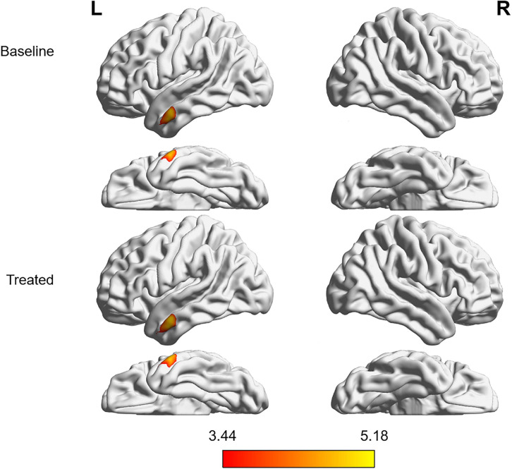
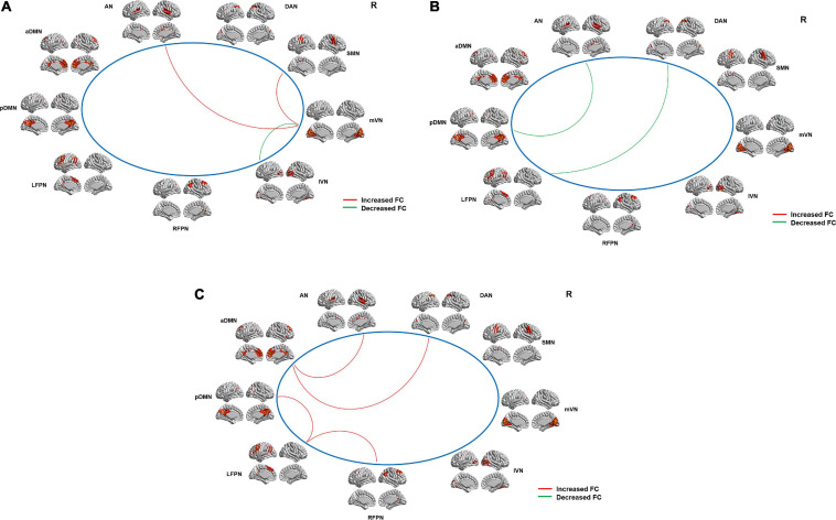
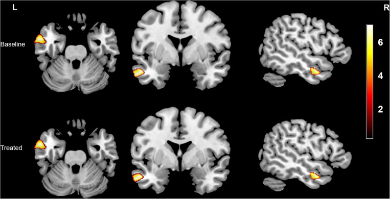
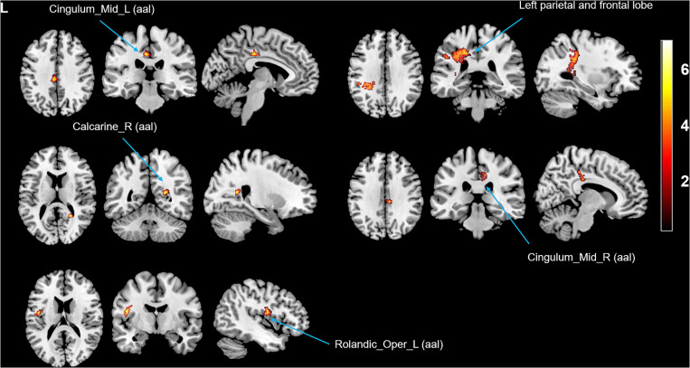
Sound therapy is one of the most common first-line treatments for idiopathic tinnitus. We aimed to investigate the brain structural and functional alterations between patients with idiopathic tinnitus without hearing loss (HL) and healthy controls (HCs) and between patients before and after sound therapy (narrow band noise). Structural and resting-state functional images were acquired from 13 tinnitus patients without HL and 18 HCs before and after 6 months of narrow band sound therapy (only patients received the treatment). Voxel-based morphometry (VBM) and independent component analysis (ICA) were conducted to separately investigate the brain structural and functional changes. Associations between brain changes and clinical variables were also performed. After the treatment, the % improvement of THI score was -1.30% (± 63.40%). Compared with HCs, tinnitus patients showed gray matter and white matter atrophy in the left middle temporal gyrus at baseline, and the gray matter volume was further reduced after the treatment. The patients also showed increased white matter volume in the cingulum (cingulate), right calcarine, left rolandic operculum, and left parietal and frontal lobes. Additionally, compared with HCs, tinnitus patients exhibited positive [medial visual network (mVN) and sensorimotor network (SMN), mVN and auditory network (AN)] and negative [mVN and lateral visual network (lVN)] internetwork functional connectivity (FC) at baseline and negative [left frontoparietal network (LFPN) and dorsal attention network (DAN), AN and posterior default mode network (pDMN)] internetwork FC after the narrow band sound therapy. The patients also showed negative [LFPN and right frontoparietal network (RFPN), LFPN and RFPN, anterior default mode network (aDMN) and AN, aDMN and DAN] internetwork FC after the treatment when compared with baseline. Our findings suggest that although the outcomes of idiopathic tinnitus patients without HL were not very good when the improvement of THI scores was used as an evaluation indicator, the patients experienced significant differences in auditory-related and non-auditory-related brain reorganization before and after the narrow band sound therapy, that is, sound therapy may have a significant effect on brain reorganization in patients with idiopathic tinnitus. This study may provide some new useful information for the understanding of mechanisms underlying idiopathic tinnitus.
声疗法是特发性耳鸣最常见的一线治疗方法之一。我们旨在研究无听力损失(HL)的特发性耳鸣患者与健康对照者(HCs)之间,以及声疗法(窄带噪声)前后患者的脑结构和功能改变。在13名无HL的耳鸣患者和18名HCs接受6个月窄带声疗法前后(仅患者接受治疗)采集结构和静息态功能图像。进行基于体素的形态学测量(VBM)和独立成分分析(ICA)以分别研究脑结构和功能变化。还进行了脑变化与临床变量之间的相关性分析。治疗后,THI评分的改善百分比为-1.30%(±63.40%)。与HCs相比,耳鸣患者在基线时左侧颞中回出现灰质和白质萎缩,治疗后灰质体积进一步减少。患者还表现出扣带(扣带回)、右侧距状裂、左侧中央沟盖以及左侧顶叶和额叶的白质体积增加。此外,与HCs相比,耳鸣患者在基线时表现出正性的[内侧视觉网络(mVN)和感觉运动网络(SMN)、mVN和听觉网络(AN)]以及负性的[mVN和外侧视觉网络(lVN)]网络间功能连接(FC),在窄带声疗法后表现出负性的[左侧额顶网络(LFPN)和背侧注意网络(DAN)、AN和后默认模式网络(pDMN)]网络间FC。与基线相比,患者在治疗后还表现出负性的[LFPN和右侧额顶网络(RFPN)、LFPN和RFPN、前默认模式网络(aDMN)和AN、aDMN和DAN]网络间FC。我们的研究结果表明,尽管以THI评分的改善作为评估指标时,无HL的特发性耳鸣患者的治疗效果不太理想,但患者在窄带声疗法前后在听觉相关和非听觉相关的脑重组方面存在显著差异,即声疗法可能对特发性耳鸣患者的脑重组有显著影响。本研究可能为理解特发性耳鸣的潜在机制提供一些新的有用信息。