• 文献检索
  • 文档翻译
  • 深度研究
  • 学术资讯
  • Suppr Zotero 插件Zotero 插件
  • 邀请有礼
  • 套餐&价格
  • 历史记录
应用&插件
Suppr Zotero 插件Zotero 插件浏览器插件Mac 客户端Windows 客户端微信小程序
定价
高级版会员购买积分包购买API积分包
服务
文献检索文档翻译深度研究API 文档MCP 服务
关于我们
关于 Suppr公司介绍联系我们用户协议隐私条款
关注我们

Suppr 超能文献

核心技术专利:CN118964589B侵权必究
粤ICP备2023148730 号-1Suppr @ 2026

文献检索

告别复杂PubMed语法,用中文像聊天一样搜索,搜遍4000万医学文献。AI智能推荐,让科研检索更轻松。

立即免费搜索

文件翻译

保留排版,准确专业,支持PDF/Word/PPT等文件格式,支持 12+语言互译。

免费翻译文档

深度研究

AI帮你快速写综述,25分钟生成高质量综述,智能提取关键信息,辅助科研写作。

立即免费体验

单侧血管搏动性耳鸣患者的频率依赖性神经活动

Frequency-Dependent Neural Activity in Patients with Unilateral Vascular Pulsatile Tinnitus.

作者信息

Lv Han, Zhao Pengfei, Liu Zhaohui, Wang Guopeng, Zeng Rong, Yan Fei, Dong Cheng, Zhang Ling, Li Rui, Wang Peng, Li Ting, Gong Shusheng, Wang Zhenchang

机构信息

Department of Radiology, Beijing Friendship Hospital, Capital Medical University, Beijing 100050, China; Neuroradiology Division, Department of Radiology, Stanford University, Stanford, CA 94305, USA.

Department of Radiology, Beijing Friendship Hospital, Capital Medical University, Beijing 100050, China.

出版信息

Neural Plast. 2016;2016:4918186. doi: 10.1155/2016/4918186. Epub 2016 Jun 20.

DOI:10.1155/2016/4918186
PMID:27413554
原文链接:https://pmc.ncbi.nlm.nih.gov/articles/PMC4931090/
Abstract

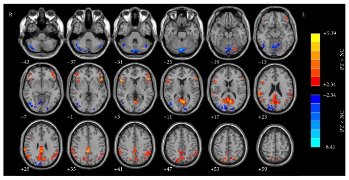
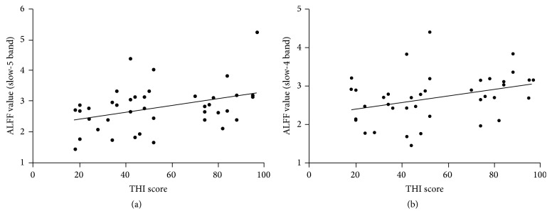
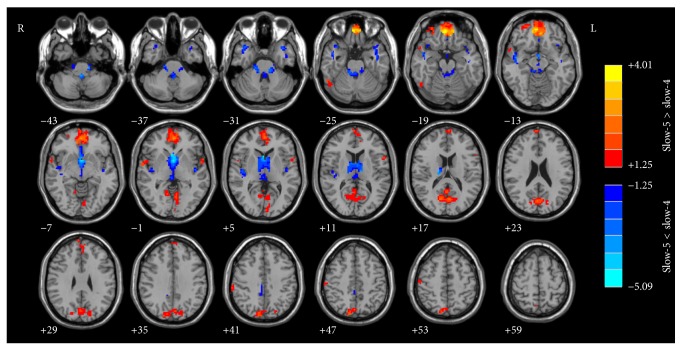
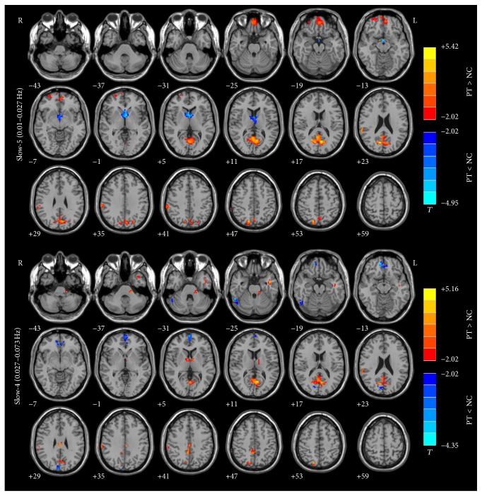
Previous resting-state functional magnetic resonance imaging (rs-fMRI) studies have shown that neurological changes are important findings in vascular pulsatile tinnitus (PT) patients. Here, we utilized rs-fMRI to measure the amplitude of low-frequency fluctuations (ALFF) in forty patients with unilateral PT and forty age-, gender-, and education-matched normal control subjects. Two different frequency bands (slow-4, 0.027-0.073 Hz, and slow-5, 0.010-0.027 Hz, which are more sensitive to subcortical and cortical neurological signal changes, resp.) were analyzed to examine the intrinsic brain activity in detail. Compared to controls, PT patients had increased ALFF values mainly in the PCu, bilateral IPL (inferior parietal lobule), left IFG (inferior frontal gyrus), and right IFG/anterior insula and decreased ALFF values in the multiple occipital areas including bilateral middle-inferior occipital lobe. For the differences of the two frequency bands, widespread ALFF differences were observed. The ALFF abnormalities in aMPFC/ACC, PCu, right IPL, and some regions of occipital and parietal cortices were greater in the slow-5 band compared to the slow-4 band. Additionally, the THI score of PT patients was positively correlated with changes in slow-5 and slow-4 band in PCu. Pulsatile tinnitus is a disease affecting the neurological activities of multiple brain regions. Slow-5 band is more sensitive in detecting the alternations. Our results also indicated the importance of pathophysiological investigations in patients with pulsatile tinnitus in the future.

摘要

以往的静息态功能磁共振成像(rs-fMRI)研究表明,神经学变化是血管搏动性耳鸣(PT)患者的重要发现。在此,我们利用rs-fMRI测量了40例单侧PT患者以及40例年龄、性别和教育程度相匹配的正常对照者的低频波动幅度(ALFF)。分析了两个不同的频段(慢4频段,0.027 - 0.073Hz,以及慢5频段,0.010 - 0.027Hz,分别对皮层下和皮层神经信号变化更敏感)以详细检查大脑的内在活动。与对照组相比,PT患者主要在楔前叶、双侧顶下小叶(IPL)、左侧额下回(IFG)以及右侧IFG/前岛叶的ALFF值增加,而在包括双侧枕叶中下部在内的多个枕叶区域ALFF值降低。对于两个频段的差异,观察到广泛的ALFF差异。与慢4频段相比,在慢5频段中,前扣带回/前扣带皮层(aMPFC/ACC)、楔前叶、右侧顶下小叶以及枕叶和顶叶皮层的一些区域的ALFF异常更为明显。此外,PT患者的耳鸣障碍指数(THI)评分与楔前叶慢5和慢4频段的变化呈正相关。搏动性耳鸣是一种影响多个脑区神经活动的疾病。慢5频段在检测这些改变方面更敏感。我们的结果还表明了未来对搏动性耳鸣患者进行病理生理学研究的重要性。

https://cdn.ncbi.nlm.nih.gov/pmc/blobs/b1df/4931090/8601f583bc78/NP2016-4918186.004.jpg
https://cdn.ncbi.nlm.nih.gov/pmc/blobs/b1df/4931090/724f28444047/NP2016-4918186.001.jpg
https://cdn.ncbi.nlm.nih.gov/pmc/blobs/b1df/4931090/efa4e823ab19/NP2016-4918186.002.jpg
https://cdn.ncbi.nlm.nih.gov/pmc/blobs/b1df/4931090/9a63f97a861b/NP2016-4918186.003.jpg
https://cdn.ncbi.nlm.nih.gov/pmc/blobs/b1df/4931090/8601f583bc78/NP2016-4918186.004.jpg

相似文献

1
Frequency-Dependent Neural Activity in Patients with Unilateral Vascular Pulsatile Tinnitus.单侧血管搏动性耳鸣患者的频率依赖性神经活动
Neural Plast. 2016;2016:4918186. doi: 10.1155/2016/4918186. Epub 2016 Jun 20.
2
Disrupted neural activity in unilateral vascular pulsatile tinnitus patients in the early stage of disease: evidence from resting-state fMRI.单侧血管搏动性耳鸣患者疾病早期的神经活动紊乱:静息态功能磁共振成像证据
Prog Neuropsychopharmacol Biol Psychiatry. 2015 Jun 3;59:91-99. doi: 10.1016/j.pnpbp.2015.01.013. Epub 2015 Jan 30.
3
Frequency-specific alternations in the amplitude of low-frequency fluctuations in chronic tinnitus.慢性耳鸣低频波动幅度的频率特异性改变
Front Neural Circuits. 2015 Oct 29;9:67. doi: 10.3389/fncir.2015.00067. eCollection 2015.
4
Frequency-dependent changes in local intrinsic oscillations in chronic primary insomnia: A study of the amplitude of low-frequency fluctuations in the resting state.慢性原发性失眠中局部内在振荡的频率依赖性变化:静息状态下低频波动幅度的研究
Neuroimage Clin. 2016 May 26;15:458-465. doi: 10.1016/j.nicl.2016.05.011. eCollection 2017.
5
Abnormal baseline brain activity in patients with pulsatile tinnitus: a resting-state FMRI study.搏动性耳鸣患者的基线脑活动异常:一项静息态 fMRI 研究。
Neural Plast. 2014;2014:549162. doi: 10.1155/2014/549162. Epub 2014 Apr 24.
6
Abnormal amplitude of low-frequency fluctuations of intrinsic brain activity in Alzheimer's disease.阿尔茨海默病中大脑内在活动低频波动的异常幅度
J Alzheimers Dis. 2014;40(2):387-97. doi: 10.3233/JAD-131322.
7
Abnormal regional activity and functional connectivity in resting-state brain networks associated with etiology confirmed unilateral pulsatile tinnitus in the early stage of disease.与病因相关的静息态脑网络中异常的区域活动和功能连接在疾病早期证实了单侧搏动性耳鸣。
Hear Res. 2017 Mar;346:55-61. doi: 10.1016/j.heares.2017.02.004. Epub 2017 Feb 7.
8
Frequency-dependent changes in the amplitude of low-frequency fluctuations in amnestic mild cognitive impairment: a resting-state fMRI study.遗忘型轻度认知障碍低频振幅的频率依赖性变化:静息态 fMRI 研究。
Neuroimage. 2011 Mar 1;55(1):287-95. doi: 10.1016/j.neuroimage.2010.11.059. Epub 2010 Nov 28.
9
Abnormal resting-state functional connectivity study in unilateral pulsatile tinnitus patients with single etiology: A seed-based functional connectivity study.单病因单侧搏动性耳鸣患者静息态功能连接异常研究:基于种子点的功能连接研究。
Eur J Radiol. 2016 Nov;85(11):2023-2029. doi: 10.1016/j.ejrad.2016.09.011. Epub 2016 Sep 13.
10
Intrinsic low-frequency oscillation changes in multiple-frequency bands in stable patients with chronic obstructive pulmonary disease.稳定期慢性阻塞性肺疾病患者多频段固有低频振荡的变化。
Brain Imaging Behav. 2021 Aug;15(4):1922-1933. doi: 10.1007/s11682-020-00385-5.

引用本文的文献

1
The quality and reliability of patient education regarding sound therapy videos for tinnitus on YouTube.YouTube上有关耳鸣声音疗法视频的患者教育的质量和可靠性。
PeerJ. 2024 Feb 1;12:e16846. doi: 10.7717/peerj.16846. eCollection 2024.
2
Altered Neurovascular Coupling in Unilateral Pulsatile Tinnitus.单侧搏动性耳鸣中神经血管耦合的改变
Front Neurosci. 2022 Jan 21;15:791436. doi: 10.3389/fnins.2021.791436. eCollection 2021.
3
Surface-Based Amplitude of Low-Frequency Fluctuation Alterations in Patients With Tinnitus Before and After Sound Therapy: A Resting-State Functional Magnetic Resonance Imaging Study.

本文引用的文献

1
Resting-state functional connectivity density mapping of etiology confirmed unilateral pulsatile tinnitus patients: Altered functional hubs in the early stage of disease.病因确诊的单侧搏动性耳鸣患者静息态功能连接密度图谱:疾病早期功能枢纽的改变
Neuroscience. 2015 Dec 3;310:27-37. doi: 10.1016/j.neuroscience.2015.09.032. Epub 2015 Sep 16.
2
Disrupted neural activity in unilateral vascular pulsatile tinnitus patients in the early stage of disease: evidence from resting-state fMRI.单侧血管搏动性耳鸣患者疾病早期的神经活动紊乱:静息态功能磁共振成像证据
Prog Neuropsychopharmacol Biol Psychiatry. 2015 Jun 3;59:91-99. doi: 10.1016/j.pnpbp.2015.01.013. Epub 2015 Jan 30.
3
耳鸣患者声音治疗前后基于表面的低频波动幅度变化:一项静息态功能磁共振成像研究
Front Neurosci. 2021 Nov 17;15:709482. doi: 10.3389/fnins.2021.709482. eCollection 2021.
4
Cortical Thickness Alterations in Patients With Tinnitus Before and After Sound Therapy: A Surface-Based Morphometry Study.耳鸣患者声音治疗前后的皮质厚度改变:一项基于表面的形态学研究。
Front Neurosci. 2021 Mar 5;15:633364. doi: 10.3389/fnins.2021.633364. eCollection 2021.
5
Altered cerebral blood flow in patients with unilateral venous pulsatile tinnitus: an arterial spin labeling study.单侧静脉性搏动性耳鸣患者脑血流改变:一项动脉自旋标记研究。
Br J Radiol. 2021 Apr 1;94(1120):20200990. doi: 10.1259/bjr.20200990. Epub 2021 Mar 18.
6
The Neural Mechanisms of Tinnitus: A Perspective From Functional Magnetic Resonance Imaging.耳鸣的神经机制:来自功能磁共振成像的视角
Front Neurosci. 2021 Feb 11;15:621145. doi: 10.3389/fnins.2021.621145. eCollection 2021.
7
Neuroanatomical Alterations in Patients With Tinnitus Before and After Sound Therapy: A Combined VBM and SCN Study.耳鸣患者声音治疗前后的神经解剖学改变:一项基于体素形态学和静息态脑网络的联合研究
Front Hum Neurosci. 2021 Jan 18;14:607452. doi: 10.3389/fnhum.2020.607452. eCollection 2020.
8
Lateralization Effects on Cerebral Blood Flow in Patients With Unilateral Pulsatile Tinnitus Measured With Arterial Spin Labeling.动脉自旋标记法测量单侧搏动性耳鸣患者脑血流的偏侧化效应
Front Hum Neurosci. 2020 Nov 17;14:591260. doi: 10.3389/fnhum.2020.591260. eCollection 2020.
9
Neuroanatomical Alterations in Patients With Tinnitus Before and After Sound Therapy: A Voxel-Based Morphometry Study.耳鸣患者声音治疗前后的神经解剖学改变:基于体素的形态学研究
Front Neurosci. 2020 Sep 8;14:911. doi: 10.3389/fnins.2020.00911. eCollection 2020.
10
Abnormal Spontaneous Neural Activity of the Central Auditory System Changes the Functional Connectivity in the Tinnitus Brain: A Resting-State Functional MRI Study.中枢听觉系统异常的自发神经活动改变耳鸣大脑中的功能连接:一项静息态功能磁共振成像研究
Front Neurosci. 2019 Dec 20;13:1314. doi: 10.3389/fnins.2019.01314. eCollection 2019.
Unilateral tinnitus: changes in connectivity and response lateralization measured with FMRI.
单侧耳鸣:通过功能磁共振成像测量的连接性和反应侧化变化。
PLoS One. 2014 Oct 20;9(10):e110704. doi: 10.1371/journal.pone.0110704. eCollection 2014.
4
Frequency-dependent neural activity in Parkinson's disease.帕金森病中频率依赖性神经活动。
Hum Brain Mapp. 2014 Dec;35(12):5815-33. doi: 10.1002/hbm.22587. Epub 2014 Jul 12.
5
Abnormal baseline brain activity in patients with pulsatile tinnitus: a resting-state FMRI study.搏动性耳鸣患者的基线脑活动异常:一项静息态 fMRI 研究。
Neural Plast. 2014;2014:549162. doi: 10.1155/2014/549162. Epub 2014 Apr 24.
6
Abnormal amplitude of low-frequency fluctuations of intrinsic brain activity in Alzheimer's disease.阿尔茨海默病中大脑内在活动低频波动的异常幅度
J Alzheimers Dis. 2014;40(2):387-97. doi: 10.3233/JAD-131322.
7
Different neural manifestations of two slow frequency bands in resting functional magnetic resonance imaging: a systemic survey at regional, interregional, and network levels.静息态功能磁共振成像中两个慢频带的不同神经表现:在区域、区间和网络水平的系统综述。
Brain Connect. 2014 May;4(4):242-55. doi: 10.1089/brain.2013.0182. Epub 2014 Mar 24.
8
Using resting state functional connectivity to unravel networks of tinnitus.利用静息态功能连接来揭示耳鸣的网络。
Hear Res. 2014 Jan;307:153-62. doi: 10.1016/j.heares.2013.07.010. Epub 2013 Jul 26.
9
Tinnitus.耳鸣。
Lancet. 2013 Nov 9;382(9904):1600-7. doi: 10.1016/S0140-6736(13)60142-7. Epub 2013 Jul 2.
10
Brain regions responsible for tinnitus distress and loudness: a resting-state FMRI study.负责耳鸣困扰和响度的大脑区域:一项静息态 fMRI 研究。
PLoS One. 2013 Jun 25;8(6):e67778. doi: 10.1371/journal.pone.0067778. Print 2013.