Saeedabadi Saghar, Abazari-Kia Amir Hossein, Rajabi Hoda, Parivar Kazem, Salehi Mohammad
Department of Biology, Faculty of Science, Islamic Azad University, Science and Research Branch, Tehran, Iran.
Department of Transgenic Animal Science, Stem Cell Technology Research Center, Tehran, Iran.
Int J Fertil Steril. 2018 Jul;12(2):157-163. doi: 10.22074/ijfs.2018.5204. Epub 2018 Mar 18.
DNA methylation is one the epigenetic mechanisms, which is critically involved in gene expression. This phenomenon is mediated by DNA methyl-transferases and is affected by environmental stress, including in vitro maturation (IVM) of oocytes. Melatonin, as an antioxidant, may theoretically be involved in epigenetic regulation via reductions of reactive oxygen species. This study was performed to investigate DNA methylation and the possibility of goat oocyte development after treatment with different concentrations of melatonin.
This experimental study was performed to investigate DNA methylation and the possibility of goat oocyte development after treatment with different concentrations of melatonin. For this purpose, oocytes with granulated cytoplasm were selected and co-cultured with at least two layers of cumulus cells in maturation medium with 10 M, 10 M, 10 M and 0-M (as control group) of melatonin. Nucleus status, glutathione content and developmental competence of the oocytes in each experimental group were assessed. Also, expression of genes associated with DNA methylation, including DNA methyltransferase 1 (DNMT1), DNA methyltransferase 3b (DNMT3b) and DNA methyltransferase 3a (DNMT3a) was evaluated by quantitative real time-polymerase chain reaction (RT-PCR).
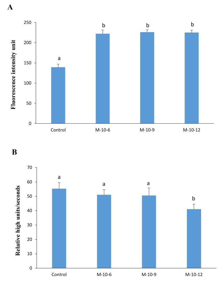
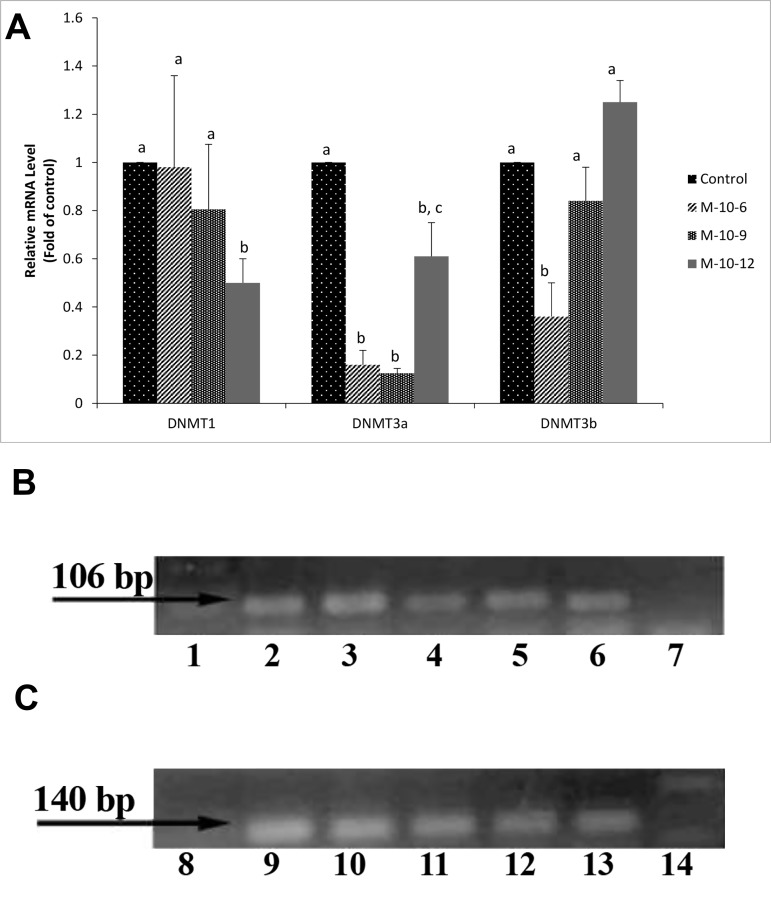
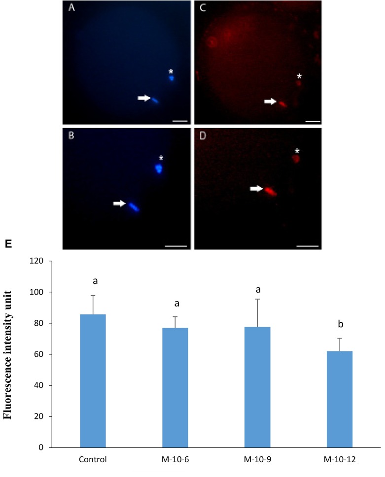
According to our findings, the percentage of oocytes that reached the M-II stage significantly increased in the 10-12 M group (P<0.05). Also, a significant elevation of glutathione content was observed in melatonin-treated oocytes (P<0.05). Analysis of blastocyst formation revealed that developmental competence of the oocytes was higher than the control group (P<0.05). It was observed that melatonin treatment decreased expression levels of DNA methyltransferases (DNMTs) and global DNA methylation (P<0.05). In addition, the expression of melatonin receptor1A (MTNR1A) was detected in both cumulus and oocyte by RT-PCR.
The results suggested that in goat model melatonin affects DNA methylation pattern, leading to an improvement in the developmental competence of the oocytes.
DNA甲基化是一种表观遗传机制,在基因表达中起关键作用。这种现象由DNA甲基转移酶介导,并受环境应激影响,包括卵母细胞的体外成熟(IVM)。褪黑素作为一种抗氧化剂,理论上可能通过减少活性氧参与表观遗传调控。本研究旨在探讨不同浓度褪黑素处理后山羊卵母细胞的DNA甲基化及发育可能性。
本实验研究旨在探讨不同浓度褪黑素处理后山羊卵母细胞的DNA甲基化及发育可能性。为此,选择细胞质颗粒化的卵母细胞,在含有10 μM、100 μM、1000 μM褪黑素和0 μM(作为对照组)的成熟培养基中与至少两层卵丘细胞共培养。评估每个实验组中卵母细胞的核状态、谷胱甘肽含量和发育能力。此外,通过定量实时聚合酶链反应(RT-PCR)评估与DNA甲基化相关基因的表达,包括DNA甲基转移酶1(DNMT1)、DNA甲基转移酶3b(DNMT3b)和DNA甲基转移酶3a(DNMT3a)。
根据我们的研究结果,10 - 12 μM组中达到M-II期的卵母细胞百分比显著增加(P<0.05)。此外,在褪黑素处理的卵母细胞中观察到谷胱甘肽含量显著升高(P<0.05)。囊胚形成分析表明,卵母细胞的发育能力高于对照组(P<0.05)。观察到褪黑素处理降低了DNA甲基转移酶(DNMTs)的表达水平和整体DNA甲基化(P<0.05)。此外,通过RT-PCR在卵丘细胞和卵母细胞中均检测到褪黑素受体1A(MTNR1A)的表达。
结果表明,在山羊模型中,褪黑素影响DNA甲基化模式,从而提高卵母细胞的发育能力。