Department of Anatomy & Neurosciences VU University Medical Center Amsterdam Neuroscience VUmc MS Center Amsterdam Amsterdam The Netherlands.
Department of Radiology Athinoula A. Martinos Center for Biomedical Imaging Massachusetts General Hospital Charlestown MA USA.
Brain Behav. 2018 Mar 30;8(5):e00954. doi: 10.1002/brb3.954. eCollection 2018 May.
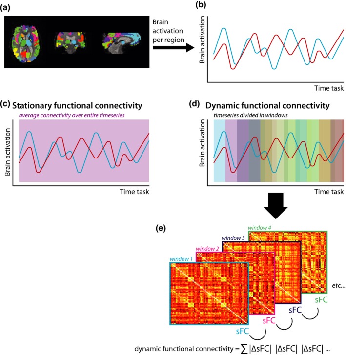
Brain dynamics (i.e., variable strength of communication between areas), even at the scale of seconds, are thought to underlie complex human behavior, such as learning and memory. In multiple sclerosis (MS), memory problems occur often and have so far only been related to "stationary" brain measures (e.g., atrophy, lesions, activation and stationary (s) functional connectivity (FC) over an entire functional scanning session). However, dynamics in FC (dFC) between the hippocampus and the (neo)cortex may be another important neurobiological substrate of memory impairment in MS that has not yet been explored. Therefore, we investigated hippocampal dFC during a functional (f) magnetic resonance imaging (MRI) episodic memory task and its relationship with verbal and visuospatial memory performance outside the MR scanner.
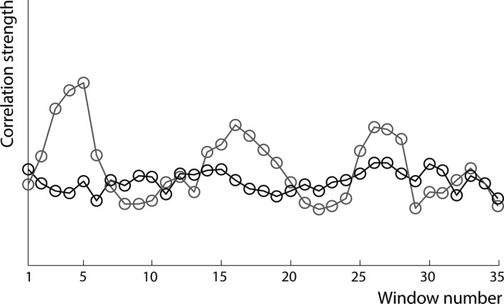
Thirty-eight MS patients and 29 healthy controls underwent neuropsychological tests to assess memory function. Imaging (1.5T) was obtained during performance of a memory task. We assessed hippocampal volume, functional activation, and sFC (i.e., FC of the hippocampus with the rest of the brain averaged over the entire scan, using an atlas-based approach). Dynamic FC of the hippocampus was calculated using a sliding window approach.
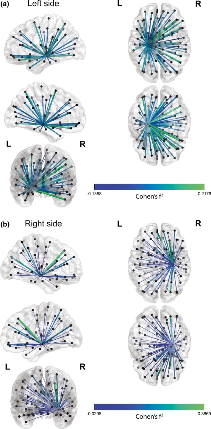
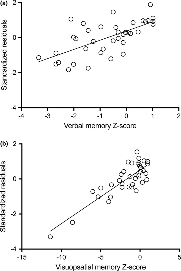
No group differences were found in hippocampal activation, sFC, and dFC. However, stepwise forward regression analyses in patients revealed that lower dFC of the left hippocampus (standardized β = -0.30; =.021) could explain an additional 7% of variance (53% in total) in verbal memory, in addition to female sex and larger left hippocampal volume. For visuospatial memory, lower dFC of the right hippocampus (standardized β = -0.38; =.013) could explain an additional 13% of variance (24% in total) in addition to higher sFC of the right hippocampus.
Low hippocampal dFC is an important indicator for maintained memory performance in MS, in addition to other hippocampal imaging measures. Hence, brain dynamics may offer new insights into the neurobiological mechanisms underlying memory (dys)function.
大脑动力学(即区域间通信强度的变化),即使在秒的尺度上,也被认为是人类复杂行为(如学习和记忆)的基础。在多发性硬化症(MS)中,经常出现记忆问题,但到目前为止,这些问题仅与“静止”的大脑测量结果相关(例如,萎缩、病变、激活以及整个功能扫描过程中的静止(s)功能连接(FC))。然而,海马体和(新)皮层之间的 FC(dFC)的动态可能是 MS 记忆障碍的另一个重要神经生物学基础,尚未得到探索。因此,我们在功能磁共振成像(fMRI)的情节记忆任务中研究了海马体的 dFC,并研究了其与磁共振扫描仪外的言语和视空间记忆表现之间的关系。
38 名 MS 患者和 29 名健康对照者接受了神经心理学测试,以评估记忆功能。成像(1.5T)是在执行记忆任务期间获得的。我们评估了海马体体积、功能激活以及 sFC(即,使用基于图谱的方法,将海马体与大脑其余部分的整个扫描平均的 FC)。使用滑动窗口方法计算了海马体的动态 FC。
患者组与对照组在海马体激活、sFC 和 dFC 方面均无差异。然而,患者的逐步正向回归分析显示,左侧海马体的 dFC 较低(标准化β=-0.30;p=.021),除了女性性别和左侧海马体体积较大外,还可以解释言语记忆的 7%的额外方差(总方差的 53%)。对于视空间记忆,右侧海马体的 dFC 较低(标准化β=-0.38;p=.013),除了右侧海马体的 sFC 较高外,还可以解释 13%的额外方差(总方差的 24%)。
除了其他海马体成像指标外,低海马体 dFC 是 MS 中记忆功能维持的重要指标。因此,大脑动力学可能为记忆(功能)障碍的神经生物学机制提供新的见解。