Department of Anatomy & Neurosciences, Neuroscience Amsterdam, VUmc MS Center Amsterdam, VU University Medical Center, Amsterdam, The Netherlands.
Department of Anatomy & Neurosciences, Neuroscience Amsterdam, VUmc MS Center Amsterdam, VU University Medical Center, Amsterdam, The Netherlands; Department of Radiology, Athinoula A. Martinos Center for Biomedical Imaging, Massachusetts General Hospital, Charlestown, MA, USA.
Neuroimage Clin. 2018 May 15;19:507-515. doi: 10.1016/j.nicl.2018.05.015. eCollection 2018.
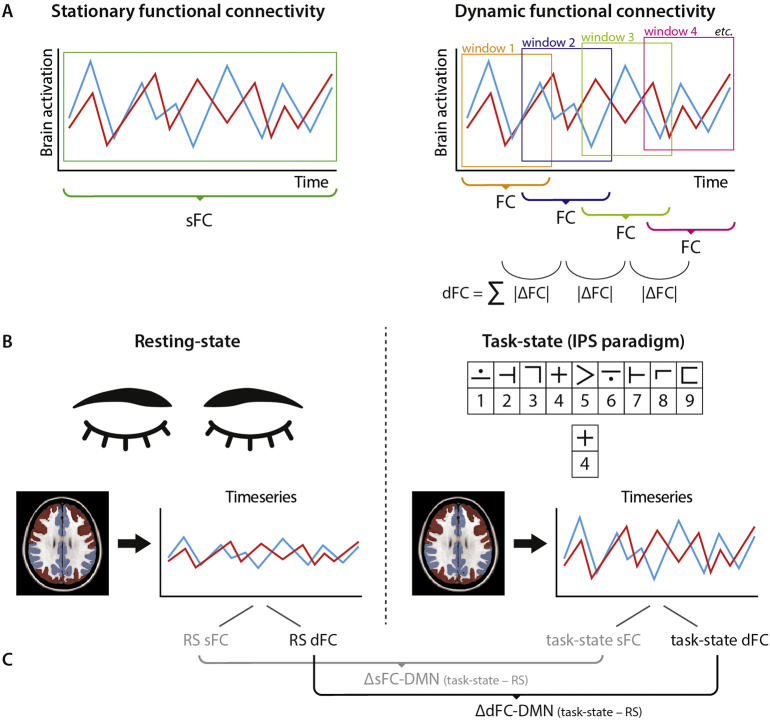
To explore the added value of dynamic functional connectivity (dFC) of the default mode network (DMN) during resting-state (RS), during an information processing speed (IPS) task, and the within-subject difference between these conditions, on top of conventional brain measures in explaining IPS in people with multiple sclerosis (pwMS).
In 29 pwMS and 18 healthy controls, IPS was assessed with the Letter Digit Substitution Test and Stroop Card I and combined into an IPS-composite score. White matter (WM), grey matter (GM) and lesion volume were measured using 3 T MRI. WM integrity was assessed with diffusion tensor imaging. During RS and task-state fMRI (i.e. symbol digit modalities task, IPS), stationary functional connectivity (sFC; average connectivity over the entire time series) and dFC (variation in connectivity using a sliding window approach) of the DMN was calculated, as well as the difference between both conditions (i.e. task-state RS; ΔsFC-DMN and ΔdFC-DMN). Regression analysis was performed to determine the most important predictors for IPS.
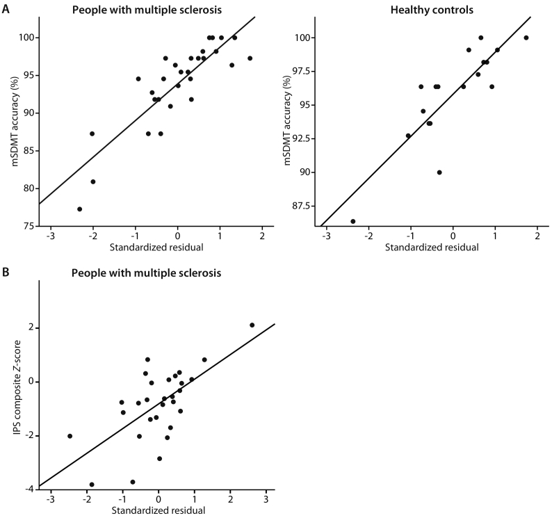
Compared to controls, pwMS performed worse on IPS-composite ( = 0.022), had lower GM volume ( < 0.05) and WM integrity ( < 0.001), but no alterations in sFC and dFC at the group level. In pwMS, 52% of variance in IPS-composite could be predicted by cortical volume (β = 0.49, = 0.01) and ΔdFC-DMN (β = 0.52, < 0.01). After adding dFC of the DMN to the model, the explained variance in IPS increased with 26% ( < 0.01).
On top of conventional brain measures, dFC from RS to task-state explains additional variance in IPS. This highlights the potential importance of the DMN to adapt upon cognitive demands to maintain intact IPS in pwMS.
在多发性硬化症(pwMS)患者中,除了常规脑测量外,静息态默认模式网络(DMN)的动态功能连接(dFC)在静息态(RS)、信息处理速度(IPS)任务期间以及这两种状态之间的个体差异,对解释 IPS 有何额外的价值。
在 29 名 pwMS 患者和 18 名健康对照者中,使用字母数字替代测试和 Stroop 卡 I 评估 IPS,并将其组合成 IPS 综合评分。使用 3T MRI 测量白质(WM)、灰质(GM)和病变体积。使用弥散张量成像评估 WM 完整性。在 RS 和任务状态 fMRI(即符号数字模态任务、IPS)期间,计算 DMN 的静息态功能连接(sFC;整个时间序列的平均连接)和 dFC(使用滑动窗口方法的连接变化),以及两种状态之间的差异(即任务状态-RS;ΔsFC-DMN 和 ΔdFC-DMN)。进行回归分析以确定 IPS 的最重要预测因子。
与对照组相比,pwMS 在 IPS 综合评分上表现更差( = 0.022),GM 体积较低( < 0.05)和 WM 完整性较低( < 0.001),但组水平的 sFC 和 dFC 没有改变。在 pwMS 中,IPS 综合评分的 52%可以通过皮质体积(β = 0.49, = 0.01)和 ΔdFC-DMN(β = 0.52, < 0.01)来预测。在将 DMN 的 dFC 添加到模型后,IPS 的解释方差增加了 26%( < 0.01)。
除了常规脑测量外,从 RS 到任务状态的 dFC 还可以解释 IPS 的额外差异。这突出表明 DMN 在认知需求下适应以维持 pwMS 中完整的 IPS 的潜在重要性。