Marr Karen, Jakimovski Dejan, Mancini Marcello, Carl Ellen, Zivadinov Robert
Buffalo Neuroimaging Analysis Center, Department of Neurology, Jacobs School of Medicine and Biomedical Sciences, University at Buffalo, State University of New York, Buffalo, New York, USA.
Institute of Biostructure and Bioimaging, National Research Council of Italy, Rome, Italy.
Ultrasound Med Biol. 2018 Aug;44(8):1762-1769. doi: 10.1016/j.ultrasmedbio.2018.04.010. Epub 2018 May 18.
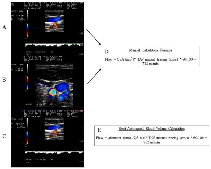
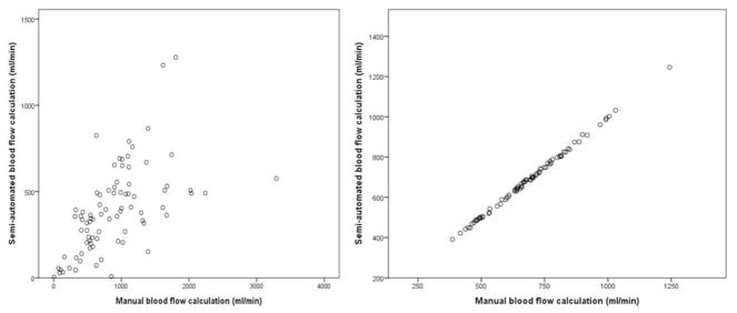
A consensus on venous flow quantification using echo spectral Doppler sonography is lacking. Doppler sonography data from 83 healthy individuals were examined using manually traced transverse cross-sectional area and diameter-derived cross-sectional area obtained in longitudinal view measurements of the internal jugular vein. Time-averaged velocity over a 4-s interval was obtained in the longitudinal plane using manual tracing of the waveform. Manual and computer-generated blood flow volume calculations were also obtained for the common carotid artery, for accuracy purposes. No differences were detected between semi-automated and manual blood flow volume calculations for the common carotid artery. The manual calculation method resulted in almost twofold larger venous internal jugular vein flow measurements compared with the semi-automated method. Doppler sonography equipment does not provide accurate automated calculation of venous size and blood flow. Until further technological development occurs, manual calculation of venous blood flow is warranted.
目前尚缺乏关于使用超声频谱多普勒超声进行静脉血流定量的共识。我们检查了83名健康个体的多普勒超声数据,采用手动追踪的横截面积以及在颈内静脉纵向视图测量中获得的直径衍生横截面积。通过手动追踪波形,在纵向平面上获得4秒间隔内的时间平均速度。为了确保准确性,还对颈总动脉进行了手动和计算机生成的血流量计算。对于颈总动脉,半自动和手动血流量计算之间未检测到差异。与半自动方法相比,手动计算方法得出的颈内静脉血流测量值几乎大两倍。多普勒超声设备无法提供准确的静脉大小和血流自动计算。在进一步的技术发展之前,有必要进行静脉血流的手动计算。