Department of Radiology, Massachusetts General Hospital, Harvard Medical School, and the Martinos Center for Biomedical Imaging, Charlestown, MA, USA; Department of Neurology, Cliniques Universitaires Saint-Luc, Institute of Neurosciences, Université Catholique de Louvain, Brussels, Belgium; Department of Neurology, Massachusetts General Hospital, Harvard Medical School, Boston, MA, USA.
Department of Biostatistics, Harvard T.H. Chan School of Public Health, Boston, MA, USA.
Alzheimers Dement. 2018 Oct;14(10):1281-1292. doi: 10.1016/j.jalz.2018.04.011. Epub 2018 May 21.
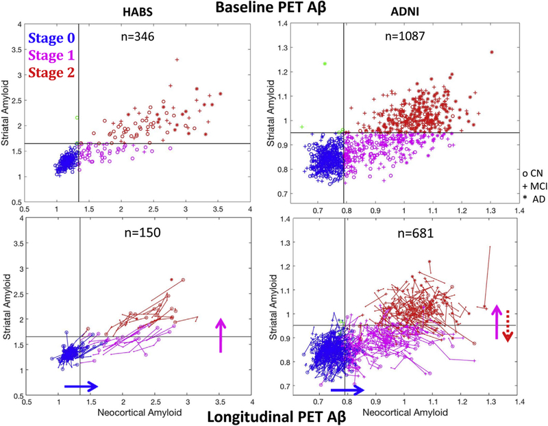
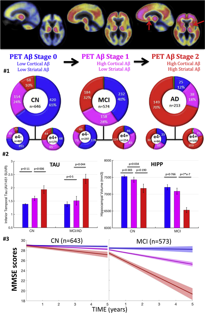
Amyloid positron emission tomography (PET) data are commonly expressed as binary measures of cortical deposition. However, not all individuals with high cortical amyloid will experience rapid cognitive decline. Motivated by postmortem data, we evaluated a three-stage PET classification: low cortical; high cortical, low striatal; and high cortical, high striatal amyloid; hypothesizing this model could better reflect Alzheimer's dementia progression than a model based only on cortical measures.
We classified PET data from 1433 participants (646 normal, 574 mild cognitive impairment, and 213 AD), explored the successive involvement of cortex and striatum using 3-year follow-up PET data, and evaluated the associations between PET stages, hippocampal volumes, and cognition.
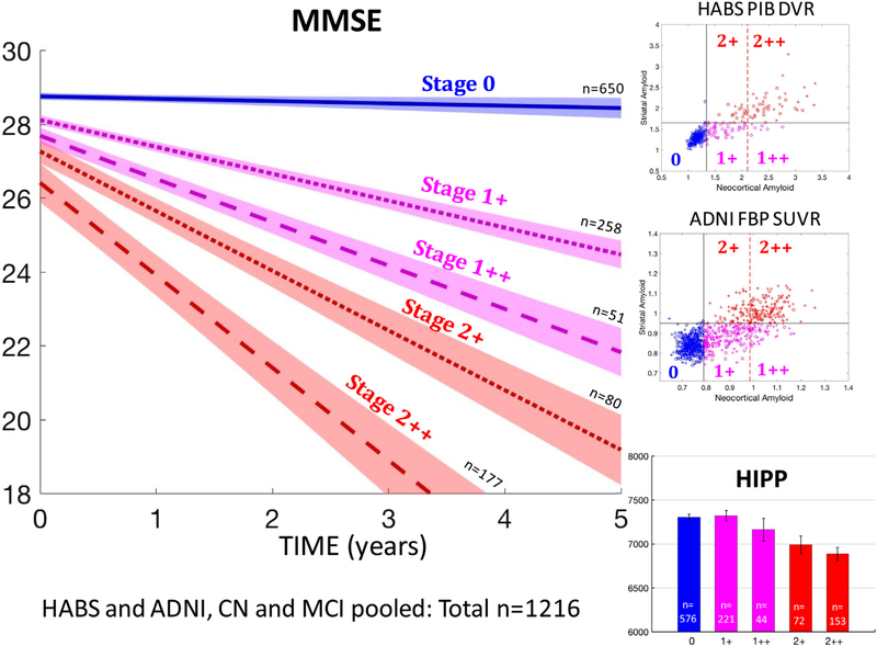
Follow-up data indicated that PET detects amyloid first in cortex and then in striatum. Our three-category staging including striatum better predicted hippocampal volumes and subsequent cognition than a three-category staging including only cortical amyloid.
PET can evaluate amyloid expansion from cortex to subcortex. Using striatal signal as a marker of advanced amyloidosis may increase predictive power in Alzheimer's dementia research.
淀粉样蛋白正电子发射断层扫描(PET)数据通常表示为皮质沉积的二进制测量值。然而,并非所有皮质高淀粉样蛋白的个体都会经历快速认知能力下降。受死后数据的启发,我们评估了 PET 的三阶段分类:低皮质;高皮质,低纹状体;高皮质,高纹状体淀粉样蛋白;假设该模型比仅基于皮质测量值的模型更能反映阿尔茨海默病的进展。
我们对 1433 名参与者(646 名正常,574 名轻度认知障碍,213 名 AD)的 PET 数据进行了分类,使用 3 年的随访 PET 数据探索皮质和纹状体的连续参与情况,并评估了 PET 分期、海马体积和认知之间的关系。
随访数据表明,PET 首先在皮质中检测到淀粉样蛋白,然后在纹状体中检测到淀粉样蛋白。我们的包括纹状体在内的三分类分期比仅包括皮质淀粉样蛋白的三分类分期更好地预测了海马体积和随后的认知。
PET 可评估从皮质到皮质下的淀粉样蛋白扩张。使用纹状体信号作为晚期淀粉样蛋白病的标志物可能会增加阿尔茨海默病研究的预测能力。