• 文献检索
  • 文档翻译
  • 深度研究
  • 学术资讯
  • Suppr Zotero 插件Zotero 插件
  • 邀请有礼
  • 套餐&价格
  • 历史记录
应用&插件
Suppr Zotero 插件Zotero 插件浏览器插件Mac 客户端Windows 客户端微信小程序
定价
高级版会员购买积分包购买API积分包
服务
文献检索文档翻译深度研究API 文档MCP 服务
关于我们
关于 Suppr公司介绍联系我们用户协议隐私条款
关注我们

Suppr 超能文献

核心技术专利:CN118964589B侵权必究
粤ICP备2023148730 号-1Suppr @ 2026

文献检索

告别复杂PubMed语法,用中文像聊天一样搜索,搜遍4000万医学文献。AI智能推荐,让科研检索更轻松。

立即免费搜索

文件翻译

保留排版,准确专业,支持PDF/Word/PPT等文件格式,支持 12+语言互译。

免费翻译文档

深度研究

AI帮你快速写综述,25分钟生成高质量综述,智能提取关键信息,辅助科研写作。

立即免费体验

缺乏 parkin 和 PINK1 会损害. 中与年龄相关的线粒体自噬。

Deficiency of parkin and PINK1 impairs age-dependent mitophagy in .

机构信息

Laboratory for Parkinson Research, Department of Neurosciences, Leuven, Belgium.

Department of Neurosciences, KU Leuven, Leuven, Belgium.

出版信息

Elife. 2018 May 29;7:e35878. doi: 10.7554/eLife.35878.

DOI:10.7554/eLife.35878
PMID:29809156
原文链接:https://pmc.ncbi.nlm.nih.gov/articles/PMC6008047/
Abstract

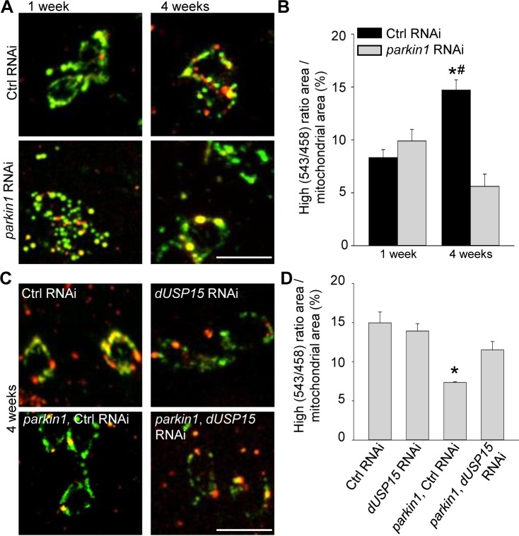
Mutations in the genes for PINK1 and parkin cause Parkinson's disease. PINK1 and parkin cooperate in the selective autophagic degradation of damaged mitochondria (mitophagy) in cultured cells. However, evidence for their role in mitophagy in vivo is still scarce. Here, we generated a model expressing the mitophagy probe mt-Keima. Using live mt-Keima imaging and correlative light and electron microscopy (CLEM), we show that mitophagy occurs in muscle cells and dopaminergic neurons in vivo, even in the absence of exogenous mitochondrial toxins. Mitophagy increases with aging, and this age-dependent rise is abrogated by PINK1 or parkin deficiency. Knockdown of the homologues of the deubiquitinases USP15 and, to a lesser extent, USP30, rescues mitophagy in the parkin-deficient flies. These data demonstrate a crucial role for parkin and PINK1 in age-dependent mitophagy in in vivo.

摘要

PINK1 和 parkin 基因突变会导致帕金森病。在培养细胞中,PINK1 和 parkin 合作进行受损线粒体的选择性自噬降解(mitophagy)。然而,它们在体内mitophagy 中的作用的证据仍然很少。在这里,我们生成了一个表达 mitophagy 探针 mt-Keima 的模型。使用活细胞 mt-Keima 成像和相关的光和电子显微镜(CLEM),我们表明mitophagy 发生在体内的肌肉细胞和多巴胺能神经元中,即使没有外源性线粒体毒素也是如此。mitophagy 随年龄增长而增加,而 PINK1 或 parkin 缺乏会消除这种年龄依赖性增加。去泛素化酶 USP15 的同源物的敲低,在较小程度上,USP30 的敲低,可挽救 parkin 缺陷果蝇中的 mitophagy。这些数据表明 parkin 和 PINK1 在体内与年龄相关的 mitophagy 中起着至关重要的作用。

https://cdn.ncbi.nlm.nih.gov/pmc/blobs/ee66/6008047/239eacdc9e9d/elife-35878-fig4.jpg
https://cdn.ncbi.nlm.nih.gov/pmc/blobs/ee66/6008047/ba425ed0c396/elife-35878-fig1.jpg
https://cdn.ncbi.nlm.nih.gov/pmc/blobs/ee66/6008047/585f8f3a3076/elife-35878-fig2.jpg
https://cdn.ncbi.nlm.nih.gov/pmc/blobs/ee66/6008047/52c6d7ebda6e/elife-35878-fig3.jpg
https://cdn.ncbi.nlm.nih.gov/pmc/blobs/ee66/6008047/239eacdc9e9d/elife-35878-fig4.jpg

相似文献

1
Deficiency of parkin and PINK1 impairs age-dependent mitophagy in .缺乏 parkin 和 PINK1 会损害. 中与年龄相关的线粒体自噬。
Elife. 2018 May 29;7:e35878. doi: 10.7554/eLife.35878.
2
Imaging mitophagy in the fruit fly.在果蝇中成像自噬体。
Autophagy. 2018;14(9):1656-1657. doi: 10.1080/15548627.2018.1496720. Epub 2018 Aug 2.
3
Assessment of mitophagy in mt-Keima revealed an essential role of the PINK1-Parkin pathway in mitophagy induction .利用 mt-Keima 评估线粒体自噬,揭示了 PINK1-Parkin 通路在诱导线粒体自噬中的重要作用。
FASEB J. 2019 Sep;33(9):9742-9751. doi: 10.1096/fj.201900073R. Epub 2019 May 23.
4
PARIS induced defects in mitochondrial biogenesis drive dopamine neuron loss under conditions of parkin or PINK1 deficiency.巴黎诱导的线粒体生物发生缺陷在 parkin 或 PINK1 缺乏的情况下驱动多巴胺神经元丢失。
Mol Neurodegener. 2020 Mar 5;15(1):17. doi: 10.1186/s13024-020-00363-x.
5
The complex I subunit NDUFA10 selectively rescues Drosophila pink1 mutants through a mechanism independent of mitophagy.复合物I亚基NDUFA10通过一种独立于线粒体自噬的机制选择性地拯救果蝇pink1突变体。
PLoS Genet. 2014 Nov 20;10(11):e1004815. doi: 10.1371/journal.pgen.1004815. eCollection 2014 Nov.
6
In vivo imaging reveals mitophagy independence in the maintenance of axonal mitochondria during normal aging.体内成像揭示了在正常衰老过程中线粒体的维持过程中线粒体自噬的独立性。
Aging Cell. 2017 Oct;16(5):1180-1190. doi: 10.1111/acel.12654. Epub 2017 Aug 7.
7
Mitochondrial CISD1/Cisd accumulation blocks mitophagy and genetic or pharmacological inhibition rescues neurodegenerative phenotypes in Pink1/parkin models.线粒体CISD1/Cisd的积累会阻断线粒体自噬,而基因或药物抑制可挽救Pink1/帕金模型中的神经退行性表型。
Mol Neurodegener. 2024 Jan 25;19(1):12. doi: 10.1186/s13024-024-00701-3.
8
Overexpression of pink1 or parkin in indirect flight muscles promotes mitochondrial proteostasis and extends lifespan in Drosophila melanogaster.过表达 pink1 或 parkin 在间接飞行肌中可促进果蝇中线粒体蛋白稳态并延长寿命。
PLoS One. 2019 Nov 12;14(11):e0225214. doi: 10.1371/journal.pone.0225214. eCollection 2019.
9
The mitochondrial deubiquitinase USP30 opposes parkin-mediated mitophagy.线粒体去泛素化酶 USP30 拮抗 parkin 介导的线粒体自噬。
Nature. 2014 Jun 19;510(7505):370-5. doi: 10.1038/nature13418. Epub 2014 Jun 4.
10
Atg1-mediated autophagy suppresses tissue degeneration in mutants by promoting mitochondrial fission in .Atg1 介导的自噬通过促进 中的线粒体裂变来抑制 突变体的组织退化。
Mol Biol Cell. 2018 Dec 15;29(26):3082-3092. doi: 10.1091/mbc.E18-04-0243. Epub 2018 Oct 24.

引用本文的文献

1
Spotlight on USP30: structure, function, disease and target inhibition.聚焦USP30:结构、功能、疾病与靶向抑制
Front Pharmacol. 2025 Aug 22;16:1629709. doi: 10.3389/fphar.2025.1629709. eCollection 2025.
2
Tumor-promoting UBR4 coordinates impaired mitophagy-associated senescence and lung adenocarcinoma pathogenesis.促进肿瘤的泛素蛋白连接酶E3组件4(UBR4)协调受损的线粒体自噬相关衰老与肺腺癌发病机制。
Proc Natl Acad Sci U S A. 2025 Jun 24;122(25):e2425015122. doi: 10.1073/pnas.2425015122. Epub 2025 Jun 18.
3
The neuroprotective role of CncC in a Drosophila model of Parkinson's disease.

本文引用的文献

1
Basal mitophagy is widespread in but minimally affected by loss of Pink1 or parkin.基础线粒体自噬在中广泛存在,但受 Pink1 或 parkin 缺失的影响最小。
J Cell Biol. 2018 May 7;217(5):1613-1622. doi: 10.1083/jcb.201801044. Epub 2018 Mar 2.
2
Mitophagy and Quality Control Mechanisms in Mitochondrial Maintenance.线粒体维持中的自噬和质量控制机制。
Curr Biol. 2018 Feb 19;28(4):R170-R185. doi: 10.1016/j.cub.2018.01.004.
3
PINK1 Phosphorylates MIC60/Mitofilin to Control Structural Plasticity of Mitochondrial Crista Junctions.PINK1 通过磷酸化 MIC60/肌球蛋白 60 来控制线粒体嵴连接的结构可塑性。
CncC在帕金森病果蝇模型中的神经保护作用。
PLoS One. 2025 May 13;20(5):e0322640. doi: 10.1371/journal.pone.0322640. eCollection 2025.
4
Attenuated PINK1 autophosphorylation play neuroprotective and anti-seizure roles in neonatal hypoxia.PINK1自磷酸化减弱在新生儿缺氧中发挥神经保护和抗癫痫作用。
Sci Rep. 2025 Apr 29;15(1):15078. doi: 10.1038/s41598-025-99915-8.
5
Antiageing strategy for neurodegenerative diseases: from mechanisms to clinical advances.神经退行性疾病的抗衰老策略:从机制到临床进展
Signal Transduct Target Ther. 2025 Mar 10;10(1):76. doi: 10.1038/s41392-025-02145-7.
6
Mitophagy is induced in human engineered heart tissue after simulated ischemia and reperfusion.在模拟缺血和再灌注后,人工程心脏组织中会诱导发生线粒体自噬。
J Cell Sci. 2025 May 1;138(9). doi: 10.1242/jcs.263408. Epub 2025 Mar 19.
7
Nutrient status alters developmental fates via a switch in mitochondrial homeodynamics.营养状况通过线粒体动态平衡的转变改变发育命运。
Nat Commun. 2025 Feb 1;16(1):1258. doi: 10.1038/s41467-025-56528-z.
8
Targeting mitophagy in neurodegenerative diseases.针对神经退行性疾病中的线粒体自噬
Nat Rev Drug Discov. 2025 Apr;24(4):276-299. doi: 10.1038/s41573-024-01105-0. Epub 2025 Jan 14.
9
The role of PINK1-Parkin in mitochondrial quality control.PTEN-induced kinase 1 (PINK1)-Parkin 通路在调控线粒体质量中的作用
Nat Cell Biol. 2024 Oct;26(10):1639-1651. doi: 10.1038/s41556-024-01513-9. Epub 2024 Oct 2.
10
Cell-Based Covalent-Capture Deubiquitinase Assay for Inhibitor Discovery.用于抑制剂发现的基于细胞的共价捕获去泛素化酶检测法
ACS Pharmacol Transl Sci. 2024 Aug 16;7(9):2827-2839. doi: 10.1021/acsptsci.4c00331. eCollection 2024 Sep 13.
Mol Cell. 2018 Mar 1;69(5):744-756.e6. doi: 10.1016/j.molcel.2018.01.026. Epub 2018 Feb 15.
4
Basal Mitophagy Occurs Independently of PINK1 in Mouse Tissues of High Metabolic Demand.基础自噬独立于 PINK1 在高代谢需求的小鼠组织中发生。
Cell Metab. 2018 Feb 6;27(2):439-449.e5. doi: 10.1016/j.cmet.2017.12.008. Epub 2018 Jan 11.
5
Cdc48 regulates a deubiquitylase cascade critical for mitochondrial fusion.Cdc48调控对线粒体融合至关重要的去泛素化酶级联反应。
Elife. 2018 Jan 8;7:e30015. doi: 10.7554/eLife.30015.
6
S-Nitrosylation of PINK1 Attenuates PINK1/Parkin-Dependent Mitophagy in hiPSC-Based Parkinson's Disease Models.PINK1 的 S-亚硝基化作用减弱了 hiPSC 帕金森病模型中 PINK1/Parkin 依赖性的线粒体自噬。
Cell Rep. 2017 Nov 21;21(8):2171-2182. doi: 10.1016/j.celrep.2017.10.068.
7
Shedding light on mitophagy in neurons: what is the evidence for PINK1/Parkin mitophagy in vivo?揭示神经元中的自噬现象:PINK1/Parkin 介导的自噬在体内的证据是什么?
Cell Mol Life Sci. 2018 Apr;75(7):1151-1162. doi: 10.1007/s00018-017-2692-9. Epub 2017 Oct 30.
8
PINK1/Parkin mitophagy and neurodegeneration-what do we really know in vivo?PINK1/帕金介导的线粒体自噬与神经退行性变——我们在体内究竟了解多少?
Curr Opin Genet Dev. 2017 Jun;44:47-53. doi: 10.1016/j.gde.2017.01.016. Epub 2017 Feb 16.
9
Atg1-independent induction of autophagy by the Drosophila Ulk3 homolog, ADUK.果蝇Ulk3同源物ADUK通过不依赖Atg1的方式诱导自噬。
FEBS J. 2016 Nov;283(21):3889-3897. doi: 10.1111/febs.13906. Epub 2016 Oct 11.
10
mito-QC illuminates mitophagy and mitochondrial architecture in vivo.线粒体质量控制(mito-QC)可在体内阐明线粒体自噬和线粒体结构。
J Cell Biol. 2016 Aug 1;214(3):333-45. doi: 10.1083/jcb.201603039. Epub 2016 Jul 25.