Department of Ophthalmology, National Taiwan University Hospital, Taipei, Taiwan.
Graduate Institute of Clinical Medicine, National Taiwan University College of Medicine, Taipei, Taiwan.
Br J Ophthalmol. 2019 Apr;103(4):511-516. doi: 10.1136/bjophthalmol-2017-311733. Epub 2018 May 29.
BACKGROUND/AIM: We investigated the microstructural changes in white matter of adults with amblyopia using diffusion spectrum imaging with systematic tract-based automatic analysis of the whole brain.
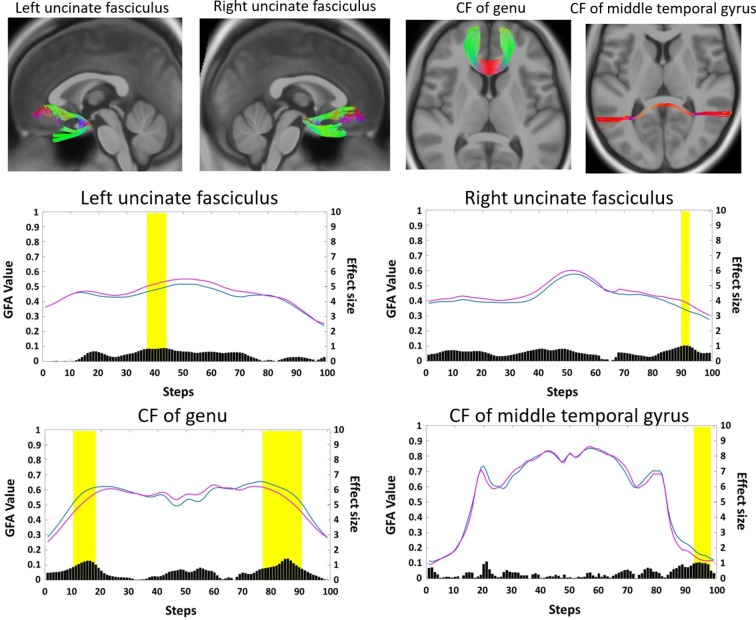
Ten adults with amblyopia (six women and four men, 33.6±10.6 years old on average) and 20 age- and sex-matched normal-sighted controls were enrolled. The mean generalised fractional anisotropy (GFA) was measured in 76 white matter tracts and compared between the experimental and control groups using a threshold-free cluster-weighted method and -test. A 2-percentile cut-off was used to identify segments with the greatest differences between the two groups.
Participants with amblyopia had significantly lower GFA values than the controls in 11 segments located in nine white matter tracts, which included the following: left arcuate fasciculus, left frontal aslant tract, left fornix and left inferior fronto-occipital fasciculus of the association fibres; left thalamic radiations of the auditory nerve and bilateral optic radiations of the projection fibres; and genu and middle temporal gyrus of the callosal fibres. Amblyopic participants had statistically higher GFA values in the bilateral uncinate fasciculus than those of the controls.
This preliminary study using whole-brain tractographic analysis of white matter reveals association between abnormal early visual processing and alterations in brain architecture, which may be related to various higher-level deficits, such as audiovisual integration and hand-eye coordination in patients with amblyopia.
背景/目的:我们使用弥散张量成像技术,对整个大脑进行基于系统的束路径自动分析,研究了弱视成年人的白质微观结构变化。
纳入 10 名弱视成年人(6 名女性,4 名男性,平均年龄 33.6±10.6 岁)和 20 名年龄和性别匹配的正常视力对照者。使用无阈值聚类加权法和 t 检验,在 76 条白质束中测量平均广义各向异性分数(GFA),并比较实验组和对照组之间的差异。采用 2%的截断值来识别两组间差异最大的节段。
与对照组相比,弱视组的 11 个节段的 GFA 值显著降低,这些节段位于 9 条白质束中,包括左侧弓状束、左侧额斜束、左侧穹窿和左侧额枕下连合纤维的联络纤维;左侧听神经丘脑辐射和双侧视放射的投射纤维;以及胼胝体的膝部和中间颞回的连合纤维。弱视组双侧钩束的 GFA 值明显高于对照组。
这项使用全脑束路径分析白质的初步研究表明,早期视觉处理异常与大脑结构改变之间存在关联,这可能与弱视患者的各种高级别缺陷有关,如视听整合和手眼协调。