Wellcome Centre for Integrative Neuroimaging, Oxford Centre for Functional Magnetic Resonance Imaging of the Brain (FMRIB), University of Oxford, UK.
Wellcome Centre for Integrative Neuroimaging, Oxford Centre for Functional Magnetic Resonance Imaging of the Brain (FMRIB), University of Oxford, UK.
Neuroimage. 2019 Jan 15;185:750-763. doi: 10.1016/j.neuroimage.2018.05.064. Epub 2018 May 28.
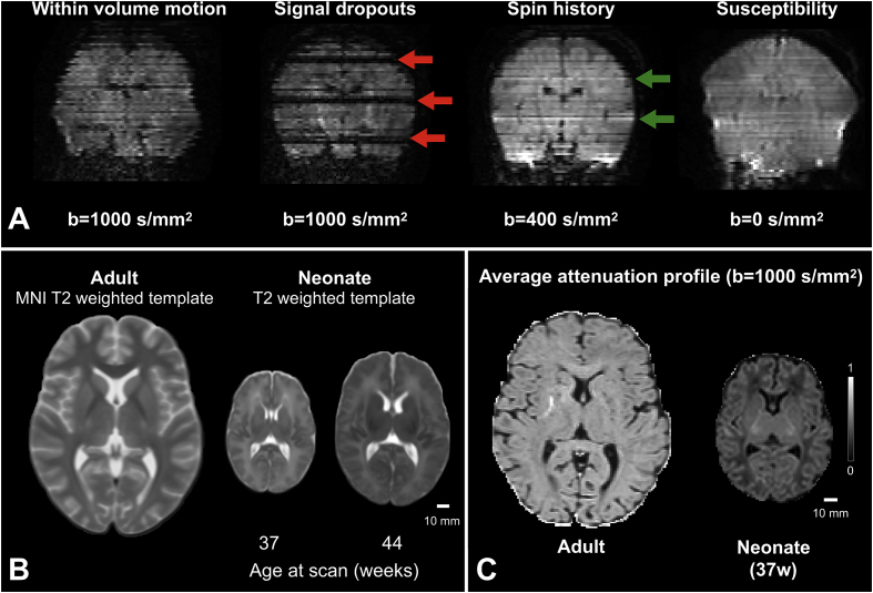
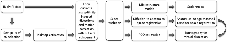
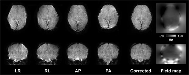
The developing Human Connectome Project is set to create and make available to the scientific community a 4-dimensional map of functional and structural cerebral connectivity from 20 to 44 weeks post-menstrual age, to allow exploration of the genetic and environmental influences on brain development, and the relation between connectivity and neurocognitive function. A large set of multi-modal MRI data from fetuses and newborn infants is currently being acquired, along with genetic, clinical and developmental information. In this overview, we describe the neonatal diffusion MRI (dMRI) image processing pipeline and the structural connectivity aspect of the project. Neonatal dMRI data poses specific challenges, and standard analysis techniques used for adult data are not directly applicable. We have developed a processing pipeline that deals directly with neonatal-specific issues, such as severe motion and motion-related artefacts, small brain sizes, high brain water content and reduced anisotropy. This pipeline allows automated analysis of in-vivo dMRI data, probes tissue microstructure, reconstructs a number of major white matter tracts, and includes an automated quality control framework that identifies processing issues or inconsistencies. We here describe the pipeline and present an exemplar analysis of data from 140 infants imaged at 38-44 weeks post-menstrual age.
正在发展的人类连接组计划将创建并向科学界提供一份从末次月经后 20 周到 44 周的大脑功能和结构连接的 4 维图谱,以探索基因和环境对大脑发育的影响,以及连接和神经认知功能之间的关系。目前正在获取大量来自胎儿和新生儿的多模态 MRI 数据,以及遗传、临床和发育信息。在这篇概述中,我们描述了新生儿弥散 MRI(dMRI)图像处理管道和项目的结构连接方面。新生儿 dMRI 数据带来了特定的挑战,而用于成人数据的标准分析技术并不直接适用。我们开发了一个处理管道,直接处理新生儿特有的问题,如严重的运动和与运动相关的伪影、小的大脑尺寸、高的脑含水量和降低的各向异性。该管道允许对体内 dMRI 数据进行自动分析,探测组织微观结构,重建多个主要的白质束,并包括一个自动质量控制框架,以识别处理问题或不一致。我们在这里描述了该管道,并展示了对 140 名在末次月经后 38-44 周成像的婴儿数据的示例分析。