Kwon Dong Rak, Park Gi-Young, Lee Sang Chul
Department of Rehabilitation Medicine, Catholic University of Daegu School of Medicine, Daegu, Republic of Korea.
Department and Research Institute of Rehabilitation Medicine, Yonsei University College of Medicine, Seoul, Republic of Korea.
Stem Cells Int. 2018 May 15;2018:7146384. doi: 10.1155/2018/7146384. eCollection 2018.
The aim of this study was to investigate regenerative effects of ultrasound- (US-) guided injection with human umbilical cord blood-derived mesenchymal stem cells (UCB-MSCs) and/or polydeoxyribonucleotide (PDRN) injection in a chronic traumatic full-thickness rotator cuff tendon tear (FTRCTT) in a rabbit model.
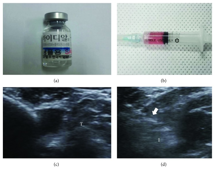
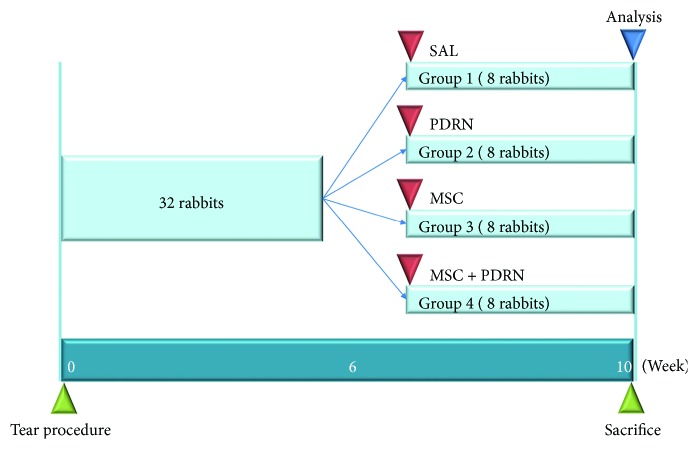
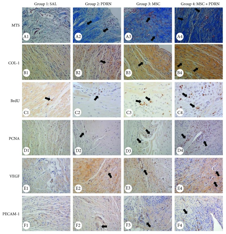
Rabbits ( = 32) were allocated into 4 groups. After a 5 mm sized FTRCTT just proximal to the insertion site on the subscapularis tendon was created by excision, the wound was immediately covered by a silicone tube to prevent natural healing. After 6 weeks, 4 injectants (0.2 mL normal saline, G1-SAL; 0.2 mL PDRN, G2-PDRN; 0.2 mL UCB-MSCs, G3-MSC; and 0.2 mL UCB-MSCs with 0.2 ml PDRN, G4-MSC + PDRN) were injected into the FTRCTT under US guidance. We evaluated gross morphologic changes on all rabbits after sacrifice. Masson's trichrome, anti-type 1 collagen antibody, bromodeoxyuridine, proliferating cell nuclear antigen, vascular endothelial growth factor, and platelet endothelial cell adhesion molecule stain were performed to evaluate histological changes. Motion analysis was also performed.
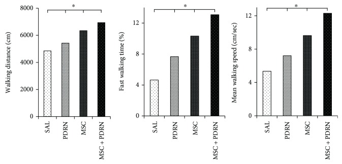
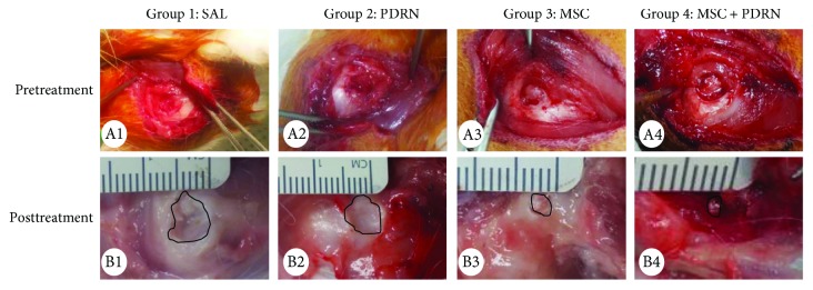
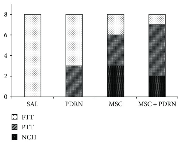
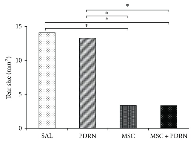
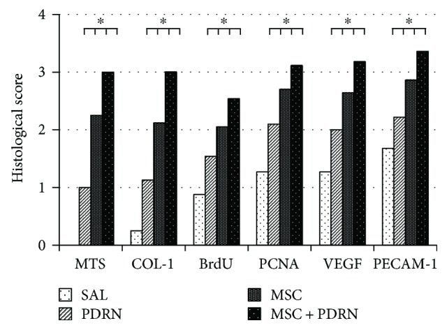
The gross morphologic mean tendon tear size in G3-MSC and G4-MSC + PDRN was significantly smaller than that in G1-SAL and G2-PDRN ( < 0.05). However, there were no significant differences in the tendon tear size between G3-MSC and G4-MSC + PDRN. In G4-MSC + PDRN, newly regenerated collagen type 1 fibers, proliferating cell activity, angiogenesis, walking distance, fast walking time, and mean walking speed were greater than those in the other three groups on histological examination and motion analysis.
Coinjection of UCB-MSCs and PDRN was more effective than UCB-MSC injection alone in histological and motion analysis in a rabbit model of chronic traumatic FTRCTT. However, there was no significant difference in gross morphologic change of tendon tear between UCB-MSCs with/without PDRN injection. The results of this study regarding the combination of UCB-MSCs and PDRN are worth additional investigations.
本研究旨在探讨超声引导下注射人脐带血间充质干细胞(UCB-MSCs)和/或聚脱氧核糖核苷酸(PDRN)对兔慢性创伤性全层肩袖肌腱撕裂(FTRCTT)模型的再生作用。
将32只兔子分为4组。通过切除在肩胛下肌腱止点近端制造一个5毫米大小的FTRCTT,伤口立即用硅胶管覆盖以防止自然愈合。6周后,在超声引导下将4种注射剂(0.2毫升生理盐水,G1-SAL;0.2毫升PDRN,G2-PDRN;0.2毫升UCB-MSCs,G3-MSC;0.2毫升UCB-MSCs加0.2毫升PDRN,G4-MSC+PDRN)注射到FTRCTT中。处死所有兔子后评估大体形态变化。进行Masson三色染色、抗I型胶原抗体染色、溴脱氧尿苷染色、增殖细胞核抗原染色、血管内皮生长因子染色和血小板内皮细胞黏附分子染色以评估组织学变化。还进行了运动分析。
G3-MSC组和G4-MSC+PDRN组的大体形态平均肌腱撕裂大小明显小于G1-SAL组和G2-PDRN组(P<0.05)。然而,G3-MSC组和G4-MSC+PDRN组之间的肌腱撕裂大小没有显著差异。在组织学检查和运动分析中,G4-MSC+PDRN组新再生的I型胶原纤维、增殖细胞活性、血管生成、行走距离、快走时间和平均行走速度均高于其他三组。
在兔慢性创伤性FTRCTT模型的组织学和运动分析中,联合注射UCB-MSCs和PDRN比单独注射UCB-MSCs更有效。然而,注射UCB-MSCs加或不加PDRN后肌腱撕裂的大体形态变化没有显著差异。本研究关于UCB-MSCs和PDRN联合应用的结果值得进一步研究。