Ogundele Olalekan M, Pardo Joaquin, Francis Joseph, Goya Rodolfo G, Lee Charles C
Department of Comparative Biomedical Sciences, School of Veterinary Medicine, Louisiana State University, Baton Rouge, LA, United States.
Institute for Biochemical Research of La Plata, School of Medicine, National University of La Plata, La Plata, Argentina.
Front Neuroanat. 2018 May 14;12:35. doi: 10.3389/fnana.2018.00035. eCollection 2018.
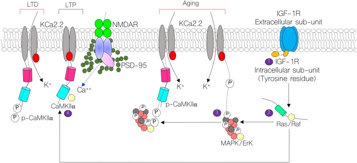
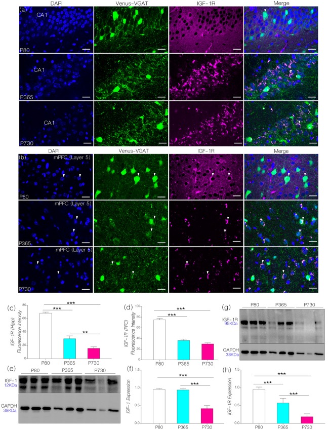
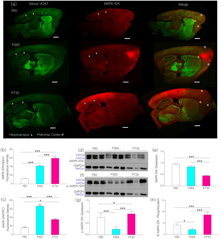
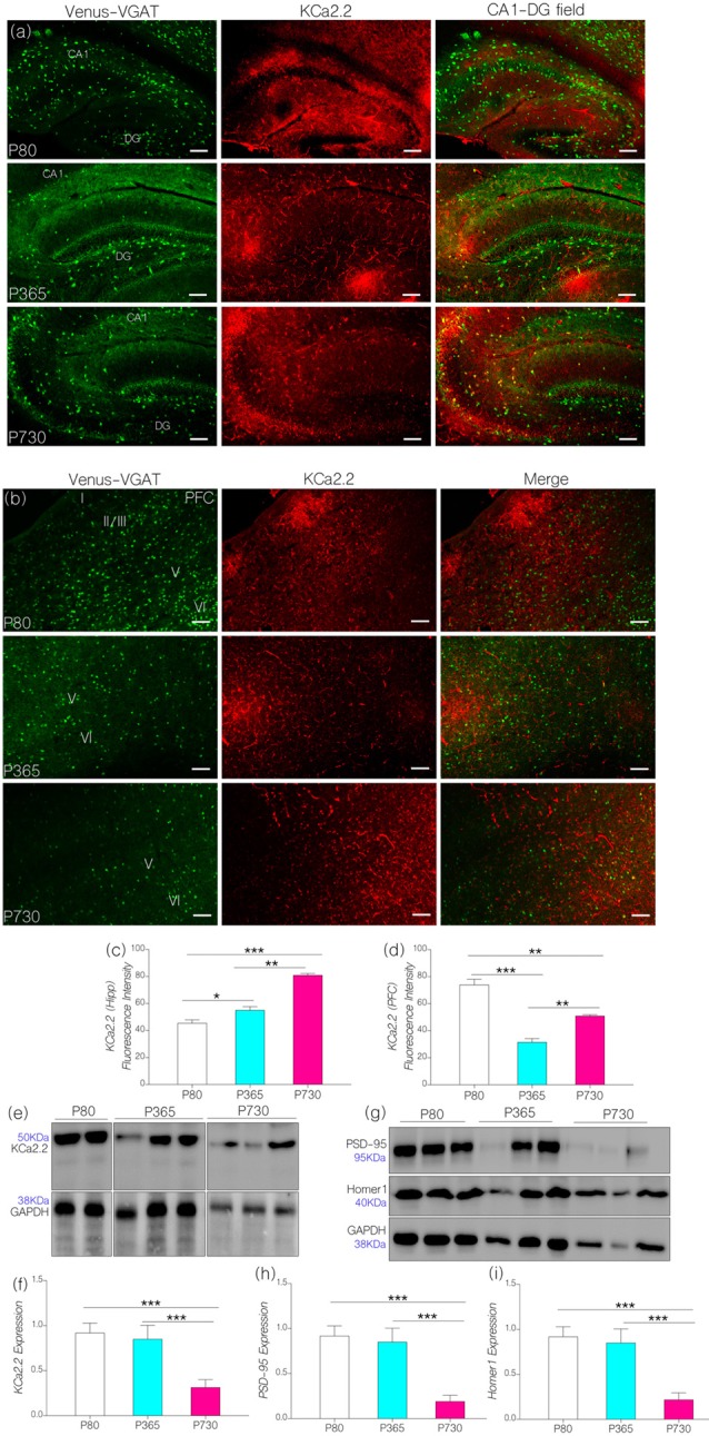
Insulin-like growth factor 1 receptor (IGF-1R) signaling regulates the activity and phosphorylation of downstream kinases linked to inflammation, neurodevelopment, aging and synaptic function. In addition to the control of Ca currents, IGF-1R signaling modulates the activity of calcium-calmodulin-dependent kinase 2 alpha (CaMKIIα) and mitogen activated protein kinase (MAPK/ErK) through multiple signaling pathways. These proteins (CaMKIIα and MAPK) regulate Ca movement and long-term potentiation (LTP). Since IGF-1R controls the synaptic activity of Ca, CaMKIIα and MAPK signaling, the possible mechanism through which an age-dependent change in IGF-1R can alter the synaptic expression and phosphorylation of these proteins in aging needs to be investigated. In this study, we evaluated the relationship between an age-dependent change in brain IGF-1R and phosphorylation of CaMKIIα/MAPK. Furthermore, we elucidated possible mechanisms through which dysregulated CaMKIIα/MAPK interaction may be linked to a change in neurotransmitter processing and synaptic function. Male C57BL/6 mice at postnatal days 80 (P80), 365 and 730 were used to study age-related neural changes in two brain regions associated with cognitive function: hippocampus and prefrontal cortex (PFC). By means of high throughput confocal imaging and quantitative immunoblotting, we evaluated the distribution and expression of IGF-1, IGF-1R, CaMKIIα, p-CaMKIIα, MAPK and p-MAPK in whole brain lysate, hippocampus and cortex. Furthermore, we compared protein expression patterns and regional changes at P80, P365 and P730. Ultimately, we determined the relative phosphorylation pattern of CaMKIIα and MAPK through quantification of neural p-CaMKIIα and p-MAPK/ErK, and IGF-1R expression for P80, P365 and P730 brain samples. In addition to a change in synaptic function, our results show a decrease in neural IGF-1/IGF-1R expression in whole brain, hippocampus and cortex of aged mice. This was associated with a significant upregulation of phosphorylated neural MAPK (p-MAPK) and decrease in total brain CaMKIIα (i.e., CaMKIIα and p-CaMKIIα) in the aged brain. Taken together, we showed that brain aging is associated with a change in neural IGF-1/IGF-1R expression and may be linked to a change in phosphorylation of synaptic kinases (CaMKIIα and MAPK) that are involved in the modulation of LTP.
胰岛素样生长因子1受体(IGF-1R)信号传导调节与炎症、神经发育、衰老和突触功能相关的下游激酶的活性和磷酸化。除了控制钙电流外,IGF-1R信号传导还通过多种信号通路调节钙/钙调蛋白依赖性激酶2α(CaMKIIα)和丝裂原活化蛋白激酶(MAPK/ErK)的活性。这些蛋白质(CaMKIIα和MAPK)调节钙的移动和长时程增强(LTP)。由于IGF-1R控制钙、CaMKIIα和MAPK信号传导的突触活性,因此需要研究IGF-1R随年龄变化而改变衰老过程中这些蛋白质的突触表达和磷酸化的可能机制。在本研究中,我们评估了脑IGF-1R随年龄变化与CaMKIIα/MAPK磷酸化之间的关系。此外,我们阐明了CaMKIIα/MAPK相互作用失调可能与神经递质加工和突触功能变化相关的可能机制。使用出生后第80天(P80)、365天和730天的雄性C57BL/6小鼠,研究与认知功能相关的两个脑区:海马体和前额叶皮质(PFC)中与年龄相关的神经变化。通过高通量共聚焦成像和定量免疫印迹,我们评估了全脑裂解物、海马体和皮质中IGF-1、IGF-1R、CaMKIIα、p-CaMKIIα、MAPK和p-MAPK的分布和表达。此外,我们比较了P80、P365和P730时的蛋白质表达模式和区域变化。最终,我们通过对P80、P365和P730脑样本中神经p-CaMKIIα和p-MAPK/ErK以及IGF-1R表达的定量,确定了CaMKIIα和MAPK的相对磷酸化模式。除了突触功能的变化外,我们的结果显示老年小鼠全脑、海马体和皮质中的神经IGF-1/IGF-1R表达降低。这与老年脑中磷酸化神经MAPK(p-MAPK)的显著上调以及全脑CaMKIIα总量(即CaMKIIα和p-CaMKIIα)的降低有关。综上所述,我们表明脑衰老与神经IGF-1/IGF-1R表达的变化有关,并且可能与参与LTP调节的突触激酶(CaMKIIα和MAPK)的磷酸化变化有关。