Department of Molecular Medicine, The Scripps Research Institute, La Jolla, CA 92037, USA.
Department of Molecular Medicine, The Scripps Research Institute, La Jolla, CA 92037, USA; Shiley Center for Orthopaedic Research and Education at Scripps Clinic, La Jolla, CA 92037, USA.
Acta Biomater. 2018 Aug;76:126-134. doi: 10.1016/j.actbio.2018.06.021. Epub 2018 Jun 14.
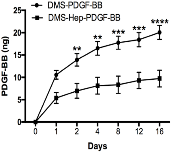
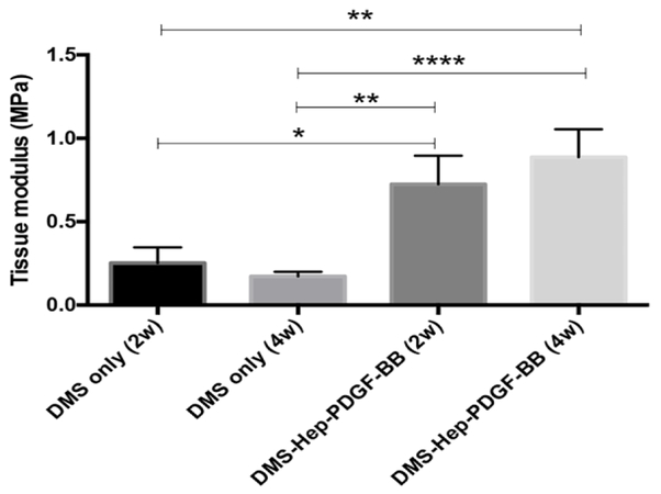
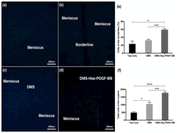
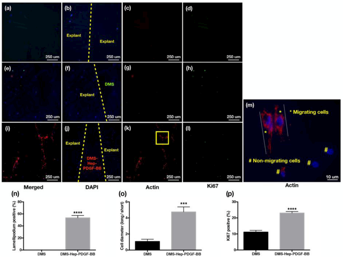
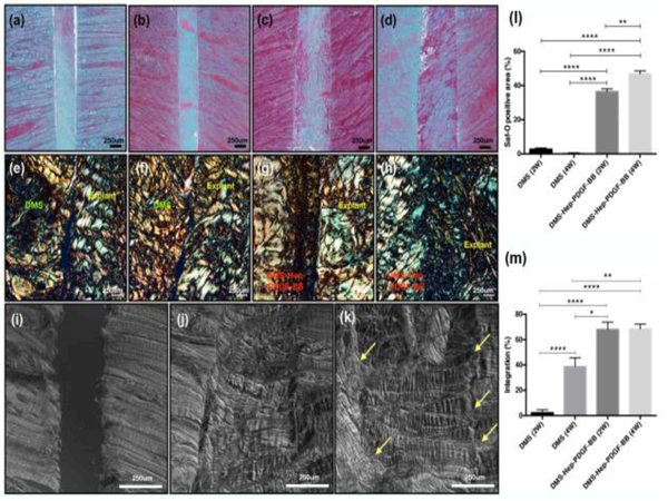
The aim of this study was to examine the potential of platelet-derived growth factor (PDGF)-coated decellularized meniscus scaffold in mediating integrative healing of meniscus tears by inducing endogenous cell migration. Fresh bovine meniscus was chemically decellularized and covalently conjugated with heparin and PDGF-BB. In vitro PDGF release kinetics was measured. The scaffold was transplanted into experimental tears in avascular bovine meniscus explants and cultured for 2 and 4 weeks. The number migrating and proliferating cells at the borderline between the scaffold and injured explant and PDGF receptor-β (PDGFRβ) expressing cells were counted. The alignment of the newly produced ECM and collagen was analyzed by Safranin-O, picrosirius red staining, and differential interference contrast (DIC). Tensile testing of the explants was performed after culture for 2 and 4 weeks. Heparin conjugated scaffold showed immobilization of high levels of PDGF-BB, with sustained release over 2 weeks. Insertion of the PDGF-BB treated scaffold in defects in avascular meniscus led to increased PDGFRβ expression, cell migration and proliferation into the defect zone. Safranin-O, picrosirius red staining and DIC showed tissue integration between the scaffold and injured explants. Tensile properties of injured explants treated with PDGF-BB coated scaffold were significantly higher than in the scaffold without PDGF. In conclusion, PDGF-BB-coated scaffold increased PDGFRβ expression and promoted migration of endogenous meniscus cells to the defect area. New matrix was formed that bridged the space between the native meniscus and the scaffold and this was associated with improved biomechanical properties. The PDGF-BB-coated scaffold will be promising for clinical translation to healing of meniscus tears.
Meniscus tears are the most common injury of the knee joint. The most prevalent forms that occur in the inner third typically do not spontaneously heal and represent a major risk factor for the development of knee osteoarthritis. The goal of this project was to develop an approach that is readily applicable for clinical use. We selected a natural and readily available decellularized meniscus scaffold and conjugated it with PDGF, which we had previously found to have strong chemotactic activity for chondrocytes and progenitor cells. The present results show that insertion of the PDGF-conjugated scaffold in defects in avascular meniscus led to endogenous cell migration and proliferation into the defect zone with tissue integration between the scaffold and injured explants and improved tensile properties. This PDGF-conjugated scaffold will be promising for a translational approach to healing of meniscus tears.
本研究旨在通过诱导内源性细胞迁移,考察血小板衍生生长因子(PDGF)涂层去细胞半月板支架在介导半月板撕裂整合愈合方面的潜力。新鲜牛半月板经化学去细胞化后,与肝素和 PDGF-BB 共价偶联。测量支架的 PDGF 释放动力学。将支架移植到无血管牛半月板外植体的实验性撕裂处,并培养 2 周和 4 周。计数支架与损伤外植体交界处以迁移和增殖的细胞数量和表达 PDGF 受体-β(PDGFRβ)的细胞数量。通过番红 O、苦味酸天狼星红染色和微分干涉对比(DIC)分析新产生的 ECM 和胶原的排列。培养 2 周和 4 周后,对外植体进行拉伸试验。肝素偶联支架显示可固定高水平的 PDGF-BB,并在 2 周内持续释放。将 PDGF-BB 处理的支架插入无血管半月板的缺损处,导致 PDGFRβ 表达增加,细胞迁移和增殖进入缺损区。番红 O、苦味酸天狼星红染色和 DIC 显示支架与损伤外植体之间的组织整合。用 PDGF-BB 涂层支架处理的损伤外植体的拉伸性能明显高于无 PDGF 的支架。结论:PDGF-BB 涂层支架增加了 PDGFRβ 的表达,并促进了内源性半月板细胞向缺损区迁移。形成了新的基质,弥合了天然半月板和支架之间的空间,这与改善生物力学性能有关。PDGF-BB 涂层支架有望在临床上用于治疗半月板撕裂。