Harvard Medical School, Boston, USA.
Harvard Medical School, Boston, USA.
Neuroimage. 2018 Oct 1;179:429-447. doi: 10.1016/j.neuroimage.2018.06.027. Epub 2018 Jun 18.
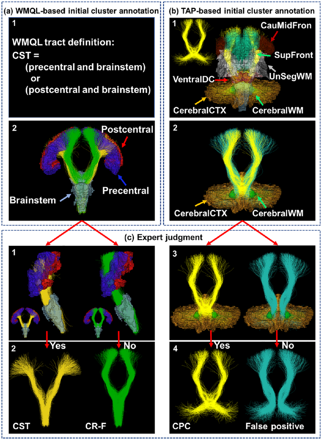
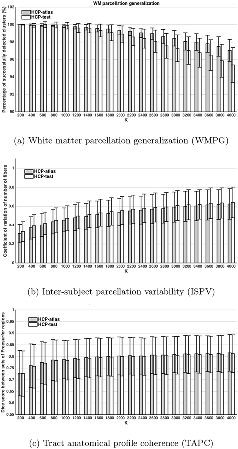
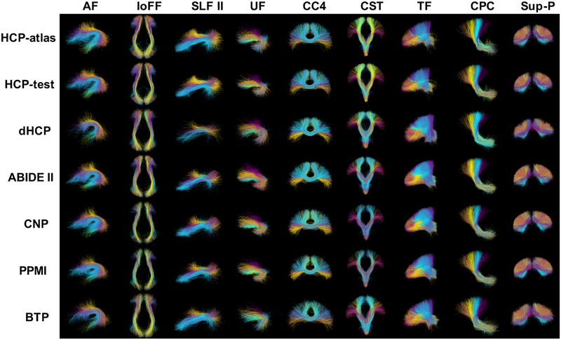
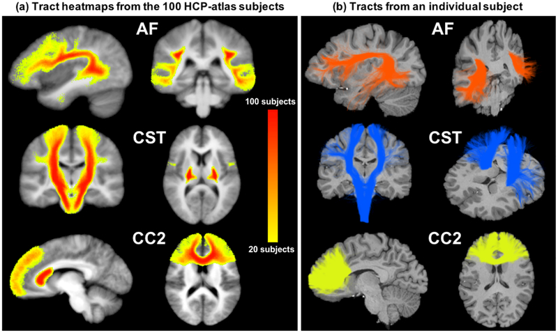
This work presents an anatomically curated white matter atlas to enable consistent white matter tract parcellation across different populations. Leveraging a well-established computational pipeline for fiber clustering, we create a tract-based white matter atlas including information from 100 subjects. A novel anatomical annotation method is proposed that leverages population-based brain anatomical information and expert neuroanatomical knowledge to annotate and categorize the fiber clusters. A total of 256 white matter structures are annotated in the proposed atlas, which provides one of the most comprehensive tract-based white matter atlases covering the entire brain to date. These structures are composed of 58 deep white matter tracts including major long range association and projection tracts, commissural tracts, and tracts related to the brainstem and cerebellar connections, plus 198 short and medium range superficial fiber clusters organized into 16 categories according to the brain lobes they connect. Potential false positive connections are annotated in the atlas to enable their exclusion from analysis or visualization. In addition, the proposed atlas allows for a whole brain white matter parcellation into 800 fiber clusters to enable whole brain connectivity analyses. The atlas and related computational tools are open-source and publicly available. We evaluate the proposed atlas using a testing dataset of 584 diffusion MRI scans from multiple independently acquired populations, across genders, the lifespan (1 day-82 years), and different health conditions (healthy control, neuropsychiatric disorders, and brain tumor patients). Experimental results show successful white matter parcellation across subjects from different populations acquired on multiple scanners, irrespective of age, gender or disease indications. Over 99% of the fiber tracts annotated in the atlas were detected in all subjects on average. One advantage in terms of robustness is that the tract-based pipeline does not require any cortical or subcortical segmentations, which can have limited success in young children and patients with brain tumors or other structural lesions. We believe this is the first demonstration of consistent automated white matter tract parcellation across the full lifespan from birth to advanced age.
这项工作提出了一个解剖学上精心策划的白质图谱,以实现不同人群之间一致的白质束分割。利用一个成熟的纤维聚类计算管道,我们创建了一个基于束的白质图谱,其中包含 100 个受试者的信息。提出了一种新的解剖注释方法,该方法利用基于人群的大脑解剖信息和专家神经解剖知识对白质束进行注释和分类。在提出的图谱中注释了总共 256 个白质结构,这是迄今为止涵盖整个大脑的最全面的基于束的白质图谱之一。这些结构由 58 个深部白质束组成,包括主要的长程联合和投射束、连合束以及与脑干和小脑连接相关的束,加上 198 个短和中程浅层纤维束,根据它们连接的脑叶分为 16 个类别。图谱中注释了潜在的假阳性连接,以便从分析或可视化中排除这些连接。此外,该图谱还允许对整个大脑的白质进行 800 个纤维束的分割,以实现整个大脑的连接分析。图谱及其相关计算工具是开源的,并且可以公开获取。我们使用来自多个独立采集的人群的 584 个弥散 MRI 扫描的测试数据集来评估所提出的图谱,这些人群跨越性别、整个生命周期(1 天至 82 岁)和不同的健康状况(健康对照、神经精神障碍和脑肿瘤患者)。实验结果表明,即使在不同年龄、性别或疾病情况下,该图谱也能在来自不同人群的多个扫描仪上成功对白质进行分割。图谱中注释的纤维束平均在所有受试者中都能检测到 99%以上。该方法具有良好的鲁棒性,其优点在于基于束的管道不需要任何皮质或皮质下分割,这在年幼的儿童和患有脑瘤或其他结构性病变的患者中可能无法成功。我们相信,这是首次在从出生到老年的整个生命周期内实现一致的自动白质束分割的演示。