Luo Xiao, Li Kaicheng, Zeng Qingze, Huang Peiyu, Jiaerken Yeerfan, Qiu Tiantian, Xu Xiaojun, Zhou Jiong, Xu Jingjing, Zhang Minming
Department of Radiology, The Second Affiliated Hospital of Zhejiang University School of Medicine, Hangzhou, China.
Department of Neurology, The Second Affiliated Hospital of Zhejiang University School of Medicine, Hangzhou, China.
Front Aging Neurosci. 2018 Jun 5;10:161. doi: 10.3389/fnagi.2018.00161. eCollection 2018.
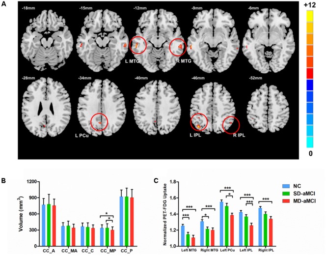
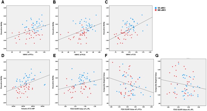
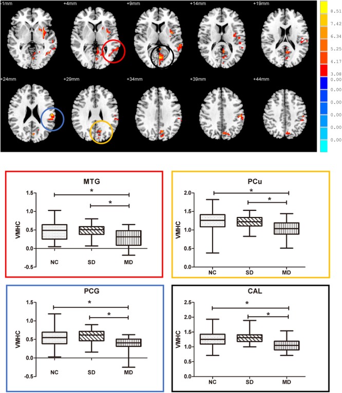
Amnestic mild cognitive impairment (aMCI) is a heterogeneous condition. Based on clinical symptoms, aMCI could be categorized into single-domain aMCI (SD-aMCI, only memory deficit) and multi-domain aMCI (MD-aMCI, one or more cognitive domain deficit). As core intrinsic functional architecture, inter-hemispheric connectivity maintains many cognitive abilities. However, few studies investigated whether SD-aMCI and MD-aMCI have different inter-hemispheric connectivity pattern. We evaluated inter-hemispheric connection pattern using fluorine-18 positron emission tomography - fluorodeoxyglucose (F PET-FDG), resting-state functional MRI and structural T1 in 49 controls, 32 SD-aMCI, and 32 MD-aMCI patients. Specifically, we analyzed the 18 PET-FDG (intensity normalized by cerebellar vermis) in a voxel-wise manner. Then, we estimated inter-hemispheric functional and structural connectivity by calculating the voxel-mirrored homotopic connectivity (VMHC) and corpus callosum (CC) subregions volume. Further, we correlated inter-hemispheric indices with the behavioral score and pathological biomarkers. We found that MD-aMCI exhibited more several inter-hemispheric connectivity damages than SD-aMCI. Specifically, MD-aMCI displayed hypometabolism in the bilateral middle temporal gyrus (MTG), inferior parietal lobe, and left precuneus (PCu) ( < 0.001, corrected). Correspondingly, MD-aMCI showed decreased VMHC in MTG, PCu, calcarine gyrus, and postcentral gyrus, as well as smaller mid-posterior CC than the SD-aMCI and controls ( < 0.05, corrected). Contrary to MD-aMCI, there were no neuroimaging indices with significant differences between SD-aMCI and controls, except reduced hypometabolism in bilateral MTG. Within aMCI patients, hypometabolism and reduced inter-hemispheric connectivity correlated with worse executive ability. Moreover, hypometabolism indices correlated to increased amyloid deposition. In conclusion, patients with MD-aMCI exhibited the more severe deficit in inter-hemispheric communication than SD-aMCI. This long-range connectivity deficit may contribute to cognitive profiles and potentially serve as a biomarker to estimate disease progression of aMCI patients.
遗忘型轻度认知障碍(aMCI)是一种异质性疾病。基于临床症状,aMCI可分为单领域aMCI(SD-aMCI,仅存在记忆缺陷)和多领域aMCI(MD-aMCI,存在一个或多个认知领域缺陷)。作为核心内在功能结构,半球间连接维持着多种认知能力。然而,很少有研究调查SD-aMCI和MD-aMCI是否具有不同的半球间连接模式。我们使用氟-18正电子发射断层扫描-氟脱氧葡萄糖(F PET-FDG)、静息态功能磁共振成像和结构T1对49名对照者、32名SD-aMCI患者和32名MD-aMCI患者的半球间连接模式进行了评估。具体而言,我们以体素方式分析了18氟代脱氧葡萄糖正电子发射断层扫描(强度经小脑蚓部标准化)。然后,我们通过计算体素镜像同伦连接性(VMHC)和胼胝体(CC)亚区域体积来估计半球间功能和结构连接性。此外,我们将半球间指标与行为评分和病理生物标志物进行了关联分析。我们发现,MD-aMCI比SD-aMCI表现出更多的半球间连接损伤。具体来说,MD-aMCI在双侧颞中回(MTG)、顶下叶和左侧楔前叶(PCu)表现出代谢减低(校正后P<0.001)。相应地,MD-aMCI在MTG、PCu、距状回和中央后回的VMHC降低,并且中后段CC比SD-aMCI和对照者更小(校正后P<0.05)。与MD-aMCI相反,除了双侧MTG代谢减低外,SD-aMCI和对照者之间没有神经影像学指标存在显著差异。在aMCI患者中,代谢减低和半球间连接性降低与更差的执行能力相关。此外,代谢减低指标与淀粉样蛋白沉积增加相关。总之,MD-aMCI患者比SD-aMCI表现出更严重的半球间交流缺陷。这种长程连接缺陷可能导致认知特征,并有可能作为评估aMCI患者疾病进展的生物标志物。