Institute of Medical Biotechnology, Department of Chemical and Biological Engineering, Friedrich-Alexander University of Erlangen-Nürnberg, Paul-Gordan-Str. 3, Erlangen, 91052, Germany.
Institute of Pharmaceutical Technology. Faculty of Biology and Pharmacy, Friedrich-Schiller-University Jena, Lessingstr. 8, Jena, 07743, Germany.
Sci Rep. 2018 Jun 20;8(1):9401. doi: 10.1038/s41598-018-27760-z.
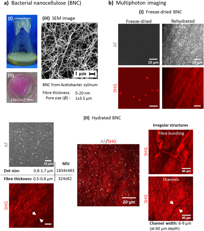
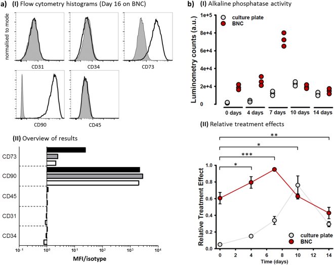
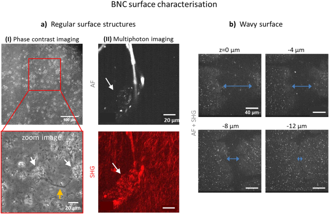
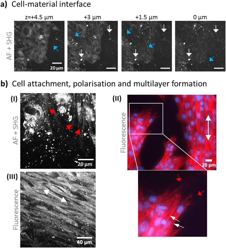
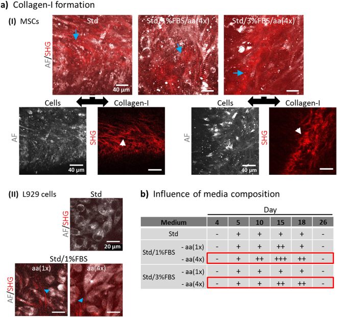
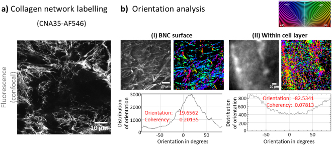
Biomimetic scaffolds are of great interest to tissue engineering (TE) and tissue repair as they support important cell functions. Scaffold coating with soluble collagen-I has been used to achieve better tissue integration in orthopaedy, however, as collagen persistence was only temporary such efforts were limited. Adequate coverage with cell-derived ECM collagen-I would promise great success, in particular for TE of mechanically challenged tissues. Here, we have used label-free, non-invasive multiphoton microscopy (MPM) to characterise bacterial nanocellulose (BNC) - a promising biomaterial for bone TE - and their potency to stimulate collagen-I formation by mesenchymal stem cells (MSCs). BNC fleeces were investigated by Second Harmonic Generation (SHG) imaging and by their characteristic autofluorescence (AF) pattern, here described for the first time. Seeded MSCs adhered fast, tight and very stable, grew to multilayers and formed characteristic, wide-spread and long-lasting collagen-I. MSCs used micron-sized lacunae and cracks on the BNC surface as cell niches. Detailed analysis using a collagen-I specific binding protein revealed a highly ordered collagen network structure at the cell-material interface. In addition, we have evidence that BNC is able to stimulate MSCs towards osteogenic differentiation. These findings offer new options for the development of engineered tissue constructs based on BNC.
仿生支架在组织工程(TE)和组织修复中非常重要,因为它们支持重要的细胞功能。支架表面涂覆可溶的胶原蛋白-I 已被用于改善矫形外科中的组织整合,但由于胶原蛋白的持久性是暂时的,因此这些努力受到限制。细胞外基质胶原蛋白-I 的充分覆盖将有望取得巨大成功,特别是对于机械挑战性组织的 TE。在这里,我们使用无标记、非侵入性的多光子显微镜(MPM)来表征细菌纳米纤维素(BNC)-一种有前途的骨 TE 生物材料-及其刺激间充质干细胞(MSCs)形成胶原蛋白-I 的能力。BNC 绒毛通过二次谐波产生(SHG)成像和其特征自体荧光(AF)模式进行了研究,这是首次对此进行描述。接种的 MSCs 快速、紧密且非常稳定地附着,生长到多层并形成特征性、广泛且持久的胶原蛋白-I。MSCs 将 BNC 表面的微米级腔隙和裂缝用作细胞龛。使用胶原蛋白-I 特异性结合蛋白进行的详细分析显示,在细胞-材料界面处存在高度有序的胶原蛋白网络结构。此外,我们有证据表明 BNC 能够刺激 MSCs 向成骨分化。这些发现为基于 BNC 的工程组织构建体的开发提供了新的选择。