Translational Research Imaging Center, Department of Clinical Radiology, University Hospital Muenster, Albert-Schweitzer-Campus 1, 48149, Muenster, Germany.
Institute for Immunology, University of Muenster, Roentgenstraße 21, 48149, Muenster, Germany.
Sci Rep. 2018 Jun 22;8(1):9563. doi: 10.1038/s41598-018-27879-z.
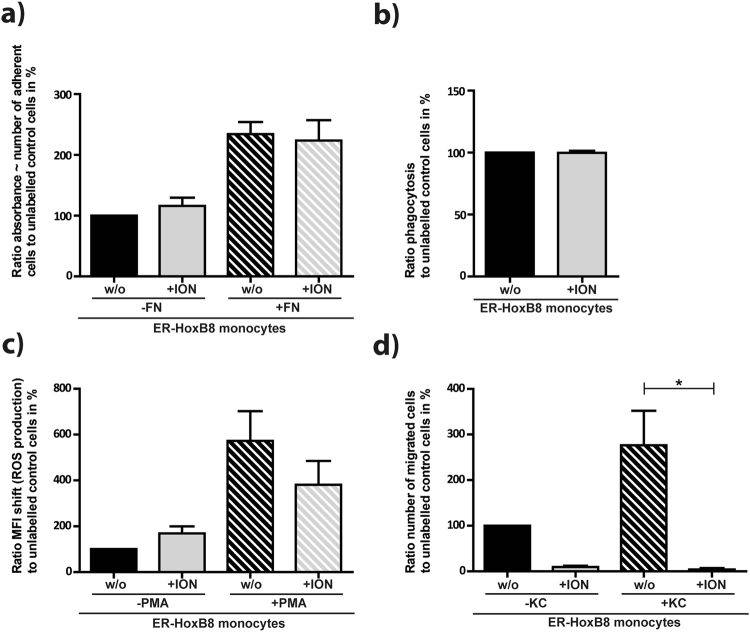
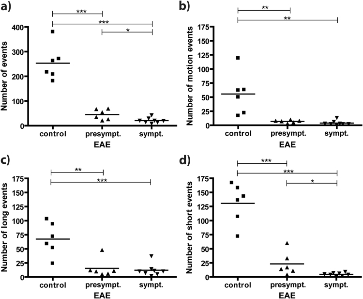
Time-lapse MRI was implemented for dynamic non-invasive cell tracking of individual slowly moving intravascular immune cells. Repetitive MRI acquisition enabled dynamic observation of iron oxide nanoparticle (ION) labelled cells. Simulations of MRI contrast indicated that only cells moving slower than 1 µm/s were detectable. Time-lapse MRI of the brain was performed after either IONs or ION-labelled monocytes were injected intravenously into naïve and experimental autoimmune encephalomyelitis (EAE) bearing mice at a presymptomatic or symptomatic stage. EAE mice showed a reduced number of slow moving, i.e. patrolling cells before and after onset of symptoms as compared to naïve controls. This observation is consistent with the notion of altered cell dynamics, i.e. higher velocities of immune cells rolling along the endothelium in the inflamed condition. Thus, time-lapse MRI enables for assessing immune cell dynamics non-invasively in deep tissue and may serve as a tool for detection or monitoring of an inflammatory response.
延时 MRI 用于对单个缓慢移动的血管内免疫细胞进行动态的非侵入性细胞追踪。重复的 MRI 采集使氧化铁纳米颗粒(ION)标记细胞的动态观察成为可能。MRI 对比模拟表明,只有移动速度慢于 1μm/s 的细胞才可以被检测到。在症状前或症状期将 ION 或 ION 标记的单核细胞静脉内注射到未受影响和实验性自身免疫性脑脊髓炎(EAE)的小鼠体内后,对大脑进行了延时 MRI。与未受影响的对照组相比,EAE 小鼠在症状出现前后表现出较慢移动的细胞(即巡逻细胞)数量减少。这种观察结果与细胞动力学改变的概念一致,即炎症状态下,免疫细胞沿内皮滚动的速度更高。因此,延时 MRI 可以非侵入性地评估深部组织中的免疫细胞动力学,并且可以作为检测或监测炎症反应的工具。