• 文献检索
  • 文档翻译
  • 深度研究
  • 学术资讯
  • Suppr Zotero 插件Zotero 插件
  • 邀请有礼
  • 套餐&价格
  • 历史记录
应用&插件
Suppr Zotero 插件Zotero 插件浏览器插件Mac 客户端Windows 客户端微信小程序
定价
高级版会员购买积分包购买API积分包
服务
文献检索文档翻译深度研究API 文档MCP 服务
关于我们
关于 Suppr公司介绍联系我们用户协议隐私条款
关注我们

Suppr 超能文献

核心技术专利:CN118964589B侵权必究
粤ICP备2023148730 号-1Suppr @ 2026

文献检索

告别复杂PubMed语法,用中文像聊天一样搜索,搜遍4000万医学文献。AI智能推荐,让科研检索更轻松。

立即免费搜索

文件翻译

保留排版,准确专业,支持PDF/Word/PPT等文件格式,支持 12+语言互译。

免费翻译文档

深度研究

AI帮你快速写综述,25分钟生成高质量综述,智能提取关键信息,辅助科研写作。

立即免费体验

利用频域光学相干断层扫描比较同胞中累及后极部的视网膜色素变性的5年病情进展:一项回顾性研究

Comparison of 5-year progression of retinitis pigmentosa involving the posterior pole among siblings by means of SD-OCT: a retrospective study.

作者信息

Colombo Leonardo, Montesano Giovanni, Sala Barbara, Patelli Fabio, Maltese Paolo, Abeshi Andi, Bertelli Matteo, Rossetti Luca

机构信息

Department of Ophthalmology, San Paolo Hospital, University of Milan, Via A. Di Rudinì 8, 20142, Milan, Italy.

Optometry and Visual Science, School of Health Sciences, City University London, London, UK.

出版信息

BMC Ophthalmol. 2018 Jun 26;18(1):153. doi: 10.1186/s12886-018-0817-z.

DOI:10.1186/s12886-018-0817-z
PMID:29940899
原文链接:https://pmc.ncbi.nlm.nih.gov/articles/PMC6019320/
Abstract

BACKGROUND

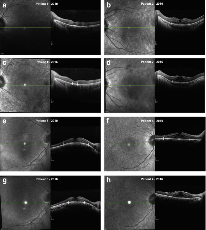
The aim of this study is to analyze and compare the progression of photoreceptor atrophy among siblings affected by retinitis pigmentosa by means of spectral SD-OCT.

METHODS

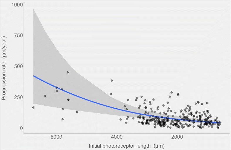
Fifty three eyes of 27 patients belonging to 12 family clusters were analyzed. To assess the annual progression rate of photoreceptor atrophy, the ellipsoid zone (EZ) line was measured in OCT sections through the fovea. We used multivariate generalized mixed effects to model the rate of progression and its relation to the initial ellipsoid zone line width.

RESULTS

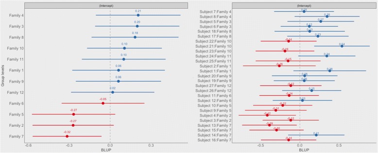
During our 4.84 years (± 1.44) mean follow up time (range 3-7) 53 eyes were examined. The ellipsoid zone line width declined with a yearly average rate of 76.4 μm (4.16% / year) (p-value < 0.0001). Progression rates were poorly correlated within family clusters (p-value = 0.23) and showed statistical difference between affected siblings (p-value = 0.007). There was no correlation between inter-familiar progression rate and mode of inheritance (p-value = 0.98) as well as between age and ellipsoid zone line width among siblings (p-value = 0.91).

CONCLUSION

RP could be extremely heterogeneous even among siblings: an accurate and sensitive method to follow the progression of the disease is fundamental for future development of clinical trials and therapy strategies.

摘要

背景

本研究旨在通过光谱频域光学相干断层扫描(SD - OCT)分析和比较患有色素性视网膜炎的兄弟姐妹中光感受器萎缩的进展情况。

方法

对来自12个家族群组的27例患者的53只眼睛进行了分析。为评估光感受器萎缩的年进展率,在通过黄斑中心凹的OCT切片中测量椭圆体带(EZ)线。我们使用多变量广义混合效应模型来模拟进展率及其与初始椭圆体带线宽度的关系。

结果

在平均4.84年(±1.44)的随访时间(范围3 - 7年)内,对53只眼睛进行了检查。椭圆体带线宽度以每年平均76.4μm(4.16%/年)的速率下降(p值<0.0001)。家族群组内的进展率相关性较差(p值 = 0.23),且患病兄弟姐妹之间存在统计学差异(p值 = 0.007)。家族间进展率与遗传模式之间(p值 = 0.98)以及兄弟姐妹的年龄与椭圆体带线宽度之间(p值 = 0.91)均无相关性。

结论

即使在兄弟姐妹之间,色素性视网膜炎也可能具有极大的异质性:一种准确且灵敏的跟踪疾病进展的方法对于未来临床试验和治疗策略的发展至关重要。

https://cdn.ncbi.nlm.nih.gov/pmc/blobs/8420/6019320/0b41444fbe85/12886_2018_817_Fig3_HTML.jpg
https://cdn.ncbi.nlm.nih.gov/pmc/blobs/8420/6019320/77486ab0b3a1/12886_2018_817_Fig1_HTML.jpg
https://cdn.ncbi.nlm.nih.gov/pmc/blobs/8420/6019320/26cf7dbfd015/12886_2018_817_Fig2_HTML.jpg
https://cdn.ncbi.nlm.nih.gov/pmc/blobs/8420/6019320/0b41444fbe85/12886_2018_817_Fig3_HTML.jpg
https://cdn.ncbi.nlm.nih.gov/pmc/blobs/8420/6019320/77486ab0b3a1/12886_2018_817_Fig1_HTML.jpg
https://cdn.ncbi.nlm.nih.gov/pmc/blobs/8420/6019320/26cf7dbfd015/12886_2018_817_Fig2_HTML.jpg
https://cdn.ncbi.nlm.nih.gov/pmc/blobs/8420/6019320/0b41444fbe85/12886_2018_817_Fig3_HTML.jpg

相似文献

1
Comparison of 5-year progression of retinitis pigmentosa involving the posterior pole among siblings by means of SD-OCT: a retrospective study.利用频域光学相干断层扫描比较同胞中累及后极部的视网膜色素变性的5年病情进展:一项回顾性研究
BMC Ophthalmol. 2018 Jun 26;18(1):153. doi: 10.1186/s12886-018-0817-z.
2
Spectral-domain optical coherence tomography measures of outer segment layer progression in patients with X-linked retinitis pigmentosa.光谱域光学相干断层扫描测量 X 连锁性视网膜炎色素变性患者的外节层进展。
JAMA Ophthalmol. 2013 Sep;131(9):1143-50. doi: 10.1001/jamaophthalmol.2013.4160.
3
Multimodal Imaging of Central Retinal Disease Progression in a 2-Year Mean Follow-up of Retinitis Pigmentosa.色素性视网膜炎2年平均随访中视网膜中央疾病进展的多模态成像
Am J Ophthalmol. 2015 Oct;160(4):786-98.e4. doi: 10.1016/j.ajo.2015.06.032. Epub 2015 Jul 9.
4
Ellipsoid zone optical intensity reduction as an early biomarker for retinitis pigmentosa.椭圆体带光强度降低可作为视网膜色素变性的早期生物标志物。
Acta Ophthalmol. 2021 Mar;99(2):e215-e221. doi: 10.1111/aos.14542. Epub 2020 Jul 23.
5
Correlation between B-scan optical coherence tomography, en face thickness map ring and hyperautofluorescent ring in retinitis pigmentosa patients.视网膜色素变性患者中B超光学相干断层扫描、表面厚度图环与高自发荧光环之间的相关性
Graefes Arch Clin Exp Ophthalmol. 2019 Aug;257(8):1601-1609. doi: 10.1007/s00417-019-04265-7. Epub 2019 May 2.
6
Outer retina analysis by optical coherence tomography in cone-rod dystrophy patients.光学相干断层扫描在外向性视网膜营养不良患者中的分析。
Retina. 2013 Oct;33(9):1877-80. doi: 10.1097/IAE.0b013e31829234e6.
7
Outer retinal layers as predictors of visual acuity in retinitis pigmentosa: a cross-sectional study.视网膜外层作为色素性视网膜炎视力的预测指标:一项横断面研究。
Graefes Arch Clin Exp Ophthalmol. 2019 Feb;257(2):265-271. doi: 10.1007/s00417-018-4185-4. Epub 2018 Nov 19.
8
Correlations between spectral-domain OCT measurements and visual acuity in cystoid macular edema associated with retinitis pigmentosa.色素性视网膜炎相关囊样黄斑水肿的光谱域 OCT 测量值与视力的相关性。
Invest Ophthalmol Vis Sci. 2013 Feb 1;54(2):1303-9. doi: 10.1167/iovs.12-10149.
9
Outer Retinal Changes Including the Ellipsoid Zone Band in Usher Syndrome 1B due to MYO7A Mutations.由于MYO7A突变导致的1B型Usher综合征的视网膜外层变化,包括椭圆体带改变
Invest Ophthalmol Vis Sci. 2016 Jul 1;57(9):OCT253-61. doi: 10.1167/iovs.15-18860.
10
Quantification of Ellipsoid Zone Changes in Retinitis Pigmentosa Using en Face Spectral Domain-Optical Coherence Tomography.使用频域光学相干断层扫描对视网膜色素变性的椭圆体带变化进行定量分析。
JAMA Ophthalmol. 2016 Jun 1;134(6):628-35. doi: 10.1001/jamaophthalmol.2016.0502.

引用本文的文献

1
Intensity-based optoretinography reveals sub-clinical deficits in cone function in retinitis pigmentosa.基于强度的视网膜电图揭示了色素性视网膜炎患者视锥细胞功能的亚临床缺陷。
Front Ophthalmol (Lausanne). 2024 Jun 4;4:1373549. doi: 10.3389/fopht.2024.1373549. eCollection 2024.
2
Short-Term Parafoveal Cone Loss Despite Preserved Ellipsoid Zone in Rod Cone Dystrophy.尽管在视杆视锥营养不良中保留了椭圆体带,但短期旁中心锥细胞丢失。
Transl Vis Sci Technol. 2021 Dec 1;10(14):11. doi: 10.1167/tvst.10.14.11.
3
Molecular Epidemiology in 591 Italian Probands With Nonsyndromic Retinitis Pigmentosa and Usher Syndrome.

本文引用的文献

1
Quantification of Ellipsoid Zone Changes in Retinitis Pigmentosa Using en Face Spectral Domain-Optical Coherence Tomography.使用频域光学相干断层扫描对视网膜色素变性的椭圆体带变化进行定量分析。
JAMA Ophthalmol. 2016 Jun 1;134(6):628-35. doi: 10.1001/jamaophthalmol.2016.0502.
2
Wide field of view swept-source optical coherence tomography for peripheral retinal disease.用于周边视网膜疾病的宽视野扫频光学相干断层扫描技术
Br J Ophthalmol. 2016 Oct;100(10):1377-82. doi: 10.1136/bjophthalmol-2015-307480. Epub 2016 Jan 11.
3
NGF eye-drops topical administration in patients with retinitis pigmentosa, a pilot study.
591 名意大利非综合征性视网膜色素变性和 Usher 综合征先证者的分子流行病学研究。
Invest Ophthalmol Vis Sci. 2021 Feb 1;62(2):13. doi: 10.1167/iovs.62.2.13.
4
Clinical characteristics and disease progression of retinitis pigmentosa associated with PDE6B mutations in Korean patients.韩国患者 PDE6B 基因突变相关视网膜炎色素变性的临床特征和疾病进展。
Sci Rep. 2020 Nov 11;10(1):19540. doi: 10.1038/s41598-020-75902-z.
5
The qualitative assessment of optical coherence tomography and the central retinal sensitivity in patients with retinitis pigmentosa.视网膜色素变性患者的光学相干断层扫描和中心视网膜敏感度的定性评估。
PLoS One. 2020 May 11;15(5):e0232700. doi: 10.1371/journal.pone.0232700. eCollection 2020.
6
Optical Coherence Tomography of Animal Models of Retinitis Pigmentosa: From Animal Studies to Clinical Applications.视网膜色素变性动物模型的光学相干断层扫描:从动物研究到临床应用。
Biomed Res Int. 2019 Oct 30;2019:8276140. doi: 10.1155/2019/8276140. eCollection 2019.
7
Determination of Length of Interdigitation Zone by Optical Coherence Tomography and Retinal Sensitivity by Microperimetry and Their Relationship to Progression of Retinitis Pigmentosa.利用光学相干断层扫描测定栉状突融合区长度和微视野计测定视网膜敏感度及其与视网膜色素变性进展的关系。
Biomed Res Int. 2019 Jun 20;2019:1217270. doi: 10.1155/2019/1217270. eCollection 2019.
神经生长因子滴眼液局部给药治疗视网膜色素变性患者的一项初步研究。
J Transl Med. 2016 Jan 9;14:8. doi: 10.1186/s12967-015-0750-3.
4
Multimodal Imaging of Central Retinal Disease Progression in a 2-Year Mean Follow-up of Retinitis Pigmentosa.色素性视网膜炎2年平均随访中视网膜中央疾病进展的多模态成像
Am J Ophthalmol. 2015 Oct;160(4):786-98.e4. doi: 10.1016/j.ajo.2015.06.032. Epub 2015 Jul 9.
5
Long-term effects of retinal gene therapy in childhood blindness.视网膜基因治疗对儿童失明的长期影响。
N Engl J Med. 2015 May 14;372(20):1954-5. doi: 10.1056/NEJMe1503419. Epub 2015 May 4.
6
Syndromic and non-syndromic forms of retinitis pigmentosa: a comprehensive Italian clinical and molecular study reveals new mutations.综合征型和非综合征型视网膜色素变性:一项全面的意大利临床和分子研究揭示了新的突变。
Genet Mol Res. 2014 Oct 27;13(4):8815-33. doi: 10.4238/2014.October.27.23.
7
A comparison of progressive loss of the ellipsoid zone (EZ) band in autosomal dominant and x-linked retinitis pigmentosa.常染色体显性遗传性视网膜色素变性与X连锁视网膜色素变性中椭圆体带(EZ)逐渐丧失的比较
Invest Ophthalmol Vis Sci. 2014 Oct 23;55(11):7417-22. doi: 10.1167/iovs.14-15013.
8
Safety evaluation of "retina implant alpha IMS"--a prospective clinical trial.“视网膜植入物阿尔法 IMS”的安全性评估——一项前瞻性临床试验。
Graefes Arch Clin Exp Ophthalmol. 2015 Mar;253(3):381-7. doi: 10.1007/s00417-014-2797-x. Epub 2014 Sep 16.
9
Recent advances of stem cell therapy for retinitis pigmentosa.视网膜色素变性干细胞治疗的最新进展
Int J Mol Sci. 2014 Aug 20;15(8):14456-74. doi: 10.3390/ijms150814456.
10
Prevalence and spatial distribution of cystoid spaces in retinitis pigmentosa: investigation with spectral domain optical coherence tomography.视网膜色素变性中囊样间隙的患病率及空间分布:光谱域光学相干断层扫描研究
Retina. 2014 May;34(5):981-8. doi: 10.1097/IAE.0000000000000010.