• 文献检索
  • 文档翻译
  • 深度研究
  • 学术资讯
  • Suppr Zotero 插件Zotero 插件
  • 邀请有礼
  • 套餐&价格
  • 历史记录
应用&插件
Suppr Zotero 插件Zotero 插件浏览器插件Mac 客户端Windows 客户端微信小程序
定价
高级版会员购买积分包购买API积分包
服务
文献检索文档翻译深度研究API 文档MCP 服务
关于我们
关于 Suppr公司介绍联系我们用户协议隐私条款
关注我们

Suppr 超能文献

核心技术专利:CN118964589B侵权必究
粤ICP备2023148730 号-1Suppr @ 2026

文献检索

告别复杂PubMed语法,用中文像聊天一样搜索,搜遍4000万医学文献。AI智能推荐,让科研检索更轻松。

立即免费搜索

文件翻译

保留排版,准确专业,支持PDF/Word/PPT等文件格式,支持 12+语言互译。

免费翻译文档

深度研究

AI帮你快速写综述,25分钟生成高质量综述,智能提取关键信息,辅助科研写作。

立即免费体验

紫外高光谱干涉显微镜。

Ultraviolet Hyperspectral Interferometric Microscopy.

机构信息

Wallace H. Coulter Department of Biomedical Engineering, Georgia Institute of Technology and Emory University, Atlanta, Georgia, USA.

Department of Pediatrics, Division of Pediatric Hematology/Oncology, Aflac Cancer Center and Blood Disorders Service of Children's Healthcare of Atlanta, Emory University School of Medicine, Atlanta, Georgia, USA.

出版信息

Sci Rep. 2018 Jul 2;8(1):9913. doi: 10.1038/s41598-018-28208-0.

DOI:10.1038/s41598-018-28208-0
PMID:29967322
原文链接:https://pmc.ncbi.nlm.nih.gov/articles/PMC6028425/
Abstract

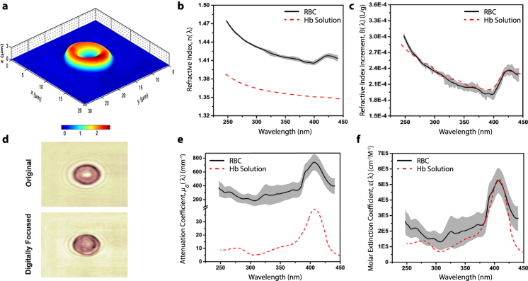
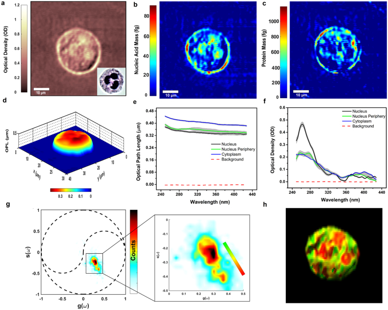
Ultraviolet (UV) spectroscopy is a powerful tool for quantitative (bio)chemical analysis, but its application to molecular imaging and microscopy has been limited. Here we introduce ultraviolet hyperspectral interferometric (UHI) microscopy, which leverages coherent detection of optical fields to overcome significant challenges associated with UV spectroscopy when applied to molecular imaging. We demonstrate that this method enables quantitative spectral analysis of important endogenous biomolecules with subcellular spatial resolution and sensitivity to nanometer-scaled structures for label-free molecular imaging of live cells.

摘要

紫外(UV)光谱学是定量(生物)化学分析的有力工具,但将其应用于分子成像和显微镜技术一直受到限制。在这里,我们引入了紫外高光谱干涉(UHI)显微镜技术,该技术利用光学场的相干检测来克服将 UV 光谱学应用于分子成像时所面临的重大挑战。我们证明,该方法能够以亚细胞空间分辨率对重要的内源性生物分子进行定量光谱分析,并对纳米级结构具有敏感性,从而实现活细胞的无标记分子成像。

https://cdn.ncbi.nlm.nih.gov/pmc/blobs/ee3c/6028425/a3771a627b8a/41598_2018_28208_Fig2_HTML.jpg
https://cdn.ncbi.nlm.nih.gov/pmc/blobs/ee3c/6028425/564bf667076d/41598_2018_28208_Fig1_HTML.jpg
https://cdn.ncbi.nlm.nih.gov/pmc/blobs/ee3c/6028425/a3771a627b8a/41598_2018_28208_Fig2_HTML.jpg
https://cdn.ncbi.nlm.nih.gov/pmc/blobs/ee3c/6028425/564bf667076d/41598_2018_28208_Fig1_HTML.jpg
https://cdn.ncbi.nlm.nih.gov/pmc/blobs/ee3c/6028425/a3771a627b8a/41598_2018_28208_Fig2_HTML.jpg

相似文献

1
Ultraviolet Hyperspectral Interferometric Microscopy.紫外高光谱干涉显微镜。
Sci Rep. 2018 Jul 2;8(1):9913. doi: 10.1038/s41598-018-28208-0.
2
Ultraviolet hyperspectral microscopy using chromatic-aberration-based iterative phase recovery.基于像散迭代相位恢复的紫外超光谱显微镜
Opt Lett. 2020 May 15;45(10):2708-2711. doi: 10.1364/OL.392634.
3
Octave-spanning hyperspectral coherent diffractive imaging in the extreme ultraviolet range.极紫外波段的倍频程超光谱相干衍射成像
Opt Express. 2015 Nov 2;23(22):28960-9. doi: 10.1364/OE.23.028960.
4
Magnified Image Spatial Spectrum (MISS) microscopy for nanometer and millisecond scale label-free imaging.用于纳米和毫秒级无标记成像的放大图像空间光谱(MISS)显微镜技术。
Opt Express. 2018 Mar 5;26(5):5423-5440. doi: 10.1364/OE.26.005423.
5
Large-aperture UV (250~400 nm) imaging spectrometer based on a solid Sagnac interferometer.基于固体萨尼亚克干涉仪的大孔径紫外(250~400纳米)成像光谱仪。
Opt Express. 2018 Dec 24;26(26):34503-34514. doi: 10.1364/OE.26.034503.
6
Efficient quantitative hyperspectral image unmixing method for large-scale Raman micro-spectroscopy data analysis.高效定量高光谱图像解混方法在大规模拉曼显微光谱数据分析中的应用。
Anal Chim Acta. 2019 Mar 7;1050:32-43. doi: 10.1016/j.aca.2018.11.018. Epub 2018 Nov 13.
7
Deep UV dispersion and absorption spectroscopy of biomolecules.生物分子的深紫外色散和吸收光谱学。
Biomed Opt Express. 2019 Jan 8;10(2):487-499. doi: 10.1364/BOE.10.000487. eCollection 2019 Feb 1.
8
Compressed sensing FTIR nano-spectroscopy and nano-imaging.压缩感知傅里叶变换红外纳米光谱学与纳米成像
Opt Express. 2018 Jul 9;26(14):18115-18124. doi: 10.1364/OE.26.018115.
9
Interferometric broadband Fourier spectroscopy with a partially coherent gas-discharge extreme ultraviolet light source.基于部分相干气体放电极紫外光源的干涉宽带傅里叶光谱学。
Opt Lett. 2015 Jun 15;40(12):2818-21. doi: 10.1364/OL.40.002818.
10
Chemical imaging by single pulse interferometric coherent anti-stokes Raman scattering microscopy.单脉冲干涉相干反斯托克斯拉曼散射显微镜的化学成像
J Phys Chem B. 2006 Mar 23;110(11):5196-204. doi: 10.1021/jp057493k.

引用本文的文献

1
Chip-based label-free incoherent super-resolution optical microscopy.基于芯片的无标记非相干超分辨率光学显微镜。
Light Sci Appl. 2025 Aug 4;14(1):259. doi: 10.1038/s41377-025-01914-x.
2
Detecting and differentiating neurotransmitters using ultraviolet plasmonic engineered native fluorescence.利用紫外等离子体工程化天然荧光检测和区分神经递质。
RSC Adv. 2023 Nov 7;13(46):32582-32588. doi: 10.1039/d3ra05405e. eCollection 2023 Oct 31.
3
Virtual Staining, Segmentation, and Classification of Blood Smears for Label-Free Hematology Analysis.

本文引用的文献

1
Is the nuclear refractive index lower than cytoplasm? Validation of phase measurements and implications for light scattering technologies.细胞核的折射率低于细胞质吗?相位测量的验证及其对光散射技术的影响。
J Biophotonics. 2017 Dec;10(12):1714-1722. doi: 10.1002/jbio.201600314. Epub 2017 Apr 18.
2
Determining the refractive index of human hemoglobin solutions by Kramers-Kronig relations with an improved absorption model.利用改进的吸收模型通过克拉默斯-克勒尼希关系测定人血红蛋白溶液的折射率。
Appl Opt. 2016 Nov 1;55(31):8951-8961. doi: 10.1364/AO.55.008951.
3
Quantitative phase imaging for medical diagnosis.
用于无标记血液学分析的血涂片虚拟染色、分割与分类
BME Front. 2022 Jul 1;2022:9853606. doi: 10.34133/2022/9853606. eCollection 2022.
4
Compact and low-cost deep-ultraviolet microscope system for label-free molecular imaging and point-of-care hematological analysis.用于无标记分子成像和即时护理血液学分析的紧凑型低成本深紫外显微镜系统。
Biomed Opt Express. 2023 Feb 24;14(3):1245-1255. doi: 10.1364/BOE.482294. eCollection 2023 Mar 1.
5
Rapid and label-free histological imaging of unprocessed surgical tissues via dark-field reflectance ultraviolet microscopy.通过暗场反射紫外显微镜对未处理手术组织进行快速且无标记的组织学成像。
iScience. 2022 Dec 21;26(1):105849. doi: 10.1016/j.isci.2022.105849. eCollection 2023 Jan 20.
6
Prostate cancer histopathology using label-free multispectral deep-UV microscopy quantifies phenotypes of tumor aggressiveness and enables multiple diagnostic virtual stains.基于无标记多光谱深紫外显微镜的前列腺癌组织病理学可定量评估肿瘤侵袭性表型,并实现多种诊断性虚拟染色。
Sci Rep. 2022 Jun 4;12(1):9329. doi: 10.1038/s41598-022-13332-9.
7
Label-free automated neutropenia detection and grading using deep-ultraviolet microscopy.使用深紫外显微镜进行无标记自动中性粒细胞减少检测和分级。
Biomed Opt Express. 2021 Sep 9;12(10):6115-6128. doi: 10.1364/BOE.434465. eCollection 2021 Oct 1.
8
Hemoglobin quantification in red blood cells via dry mass mapping based on UV absorption.基于紫外吸收的干物质测绘法对红细胞内血红蛋白进行定量。
J Biomed Opt. 2021 Aug;26(8). doi: 10.1117/1.JBO.26.8.086501.
9
Label-free imaging and classification of live P. falciparum enables high performance parasitemia quantification without fixation or staining.无标记成像和分类活的恶性疟原虫,可在不固定或染色的情况下实现高性能的疟原虫定量。
PLoS Comput Biol. 2021 Aug 9;17(8):e1009257. doi: 10.1371/journal.pcbi.1009257. eCollection 2021 Aug.
10
Label-free hematology analysis using deep-ultraviolet microscopy.无标记血液学分析的深紫外显微镜方法。
Proc Natl Acad Sci U S A. 2020 Jun 30;117(26):14779-14789. doi: 10.1073/pnas.2001404117. Epub 2020 Jun 19.
用于医学诊断的定量相位成像。
J Biophotonics. 2017 Feb;10(2):177-205. doi: 10.1002/jbio.201600113. Epub 2016 Aug 19.
4
Influence of defocus on quantitative analysis of microscopic objects and individual cells with digital holography.离焦对数字全息术用于微观物体和单个细胞定量分析的影响。
Biomed Opt Express. 2015 May 11;6(6):2067-75. doi: 10.1364/BOE.6.002067. eCollection 2015 Jun 1.
5
Spectro-refractometry of individual microscopic objects using swept-source quantitative phase imaging.使用扫频源定量相位成像对单个微观物体进行光谱折射分析。
Anal Chem. 2013 Nov 5;85(21):10519-25. doi: 10.1021/ac402521u. Epub 2013 Oct 17.
6
Intracellular protein and nucleic acid measured in eight cell types using deep-ultraviolet mass mapping.使用深紫外质谱映射技术在 8 种细胞类型中测量细胞内蛋白质和核酸。
Cytometry A. 2013 Jun;83(6):540-51. doi: 10.1002/cyto.a.22277. Epub 2013 Mar 15.
7
Quantitative phase spectroscopy.定量相位光谱学。
Biomed Opt Express. 2012 May 1;3(5):958-65. doi: 10.1364/BOE.3.000958. Epub 2012 Apr 12.
8
Nonlinear phase dispersion spectroscopy.非线性相位色散光谱学。
Opt Lett. 2011 Dec 1;36(23):4665-7. doi: 10.1364/OL.36.004665.
9
Deep ultraviolet mapping of intracellular protein and nucleic acid in femtograms per pixel.皮微微升每像素水平下细胞内蛋白质和核酸的深紫外成像。
Cytometry A. 2011 Nov;79(11):920-32. doi: 10.1002/cyto.a.21111. Epub 2011 Jul 27.
10
Inhibition of the chlorinating activity of myeloperoxidase by tempol: revisiting the kinetics and mechanisms.次氯酸根酶(MPO)的氯化活性被四甲基哌啶氧化物(Tempol)抑制:动力学和机制的再探讨。
Biochem J. 2011 Nov 1;439(3):423-31. doi: 10.1042/BJ20110555.