• 文献检索
  • 文档翻译
  • 深度研究
  • 学术资讯
  • Suppr Zotero 插件Zotero 插件
  • 邀请有礼
  • 套餐&价格
  • 历史记录
应用&插件
Suppr Zotero 插件Zotero 插件浏览器插件Mac 客户端Windows 客户端微信小程序
定价
高级版会员购买积分包购买API积分包
服务
文献检索文档翻译深度研究API 文档MCP 服务
关于我们
关于 Suppr公司介绍联系我们用户协议隐私条款
关注我们

Suppr 超能文献

核心技术专利:CN118964589B侵权必究
粤ICP备2023148730 号-1Suppr @ 2026

文献检索

告别复杂PubMed语法,用中文像聊天一样搜索,搜遍4000万医学文献。AI智能推荐,让科研检索更轻松。

立即免费搜索

文件翻译

保留排版,准确专业,支持PDF/Word/PPT等文件格式,支持 12+语言互译。

免费翻译文档

深度研究

AI帮你快速写综述,25分钟生成高质量综述,智能提取关键信息,辅助科研写作。

立即免费体验

使用心脏 MRI 中的室间隔角识别合并毛细血管前和毛细血管后肺动脉高压的高危患者。

Identifying At-Risk Patients with Combined Pre- and Postcapillary Pulmonary Hypertension Using Interventricular Septal Angle at Cardiac MRI.

机构信息

From the Academic Department of Radiology, Academic Unit of Radiology, Department of Infection, Immunity & Cardiovascular Disease, Magnetic Resonance Imaging Unit, University of Sheffield, Royal Hallamshire Hospital, Glossop Rd, Floor C, Sheffield S10 2JF, England (C.S.J., J.M.W., E.T., D.C., A.J.S.); Sheffield Pulmonary Vascular Disease Institute (C.E., R.C., A.C., D.G.K.) and Department of Radiology (S.R.), Sheffield Teaching Hospitals, Sheffield, England; and Insigneo Institute for In Silico Medicine, University of Sheffield, Sheffield, England (A.J.S.).

出版信息

Radiology. 2018 Oct;289(1):61-68. doi: 10.1148/radiol.2018180120. Epub 2018 Jul 3.

DOI:10.1148/radiol.2018180120
PMID:29969067
原文链接:https://pmc.ncbi.nlm.nih.gov/articles/PMC6190488/
Abstract

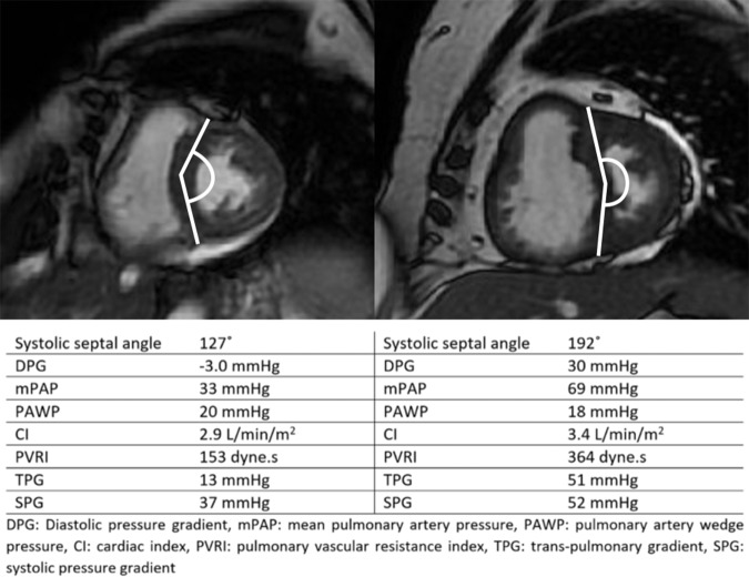
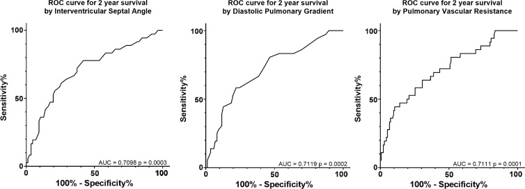
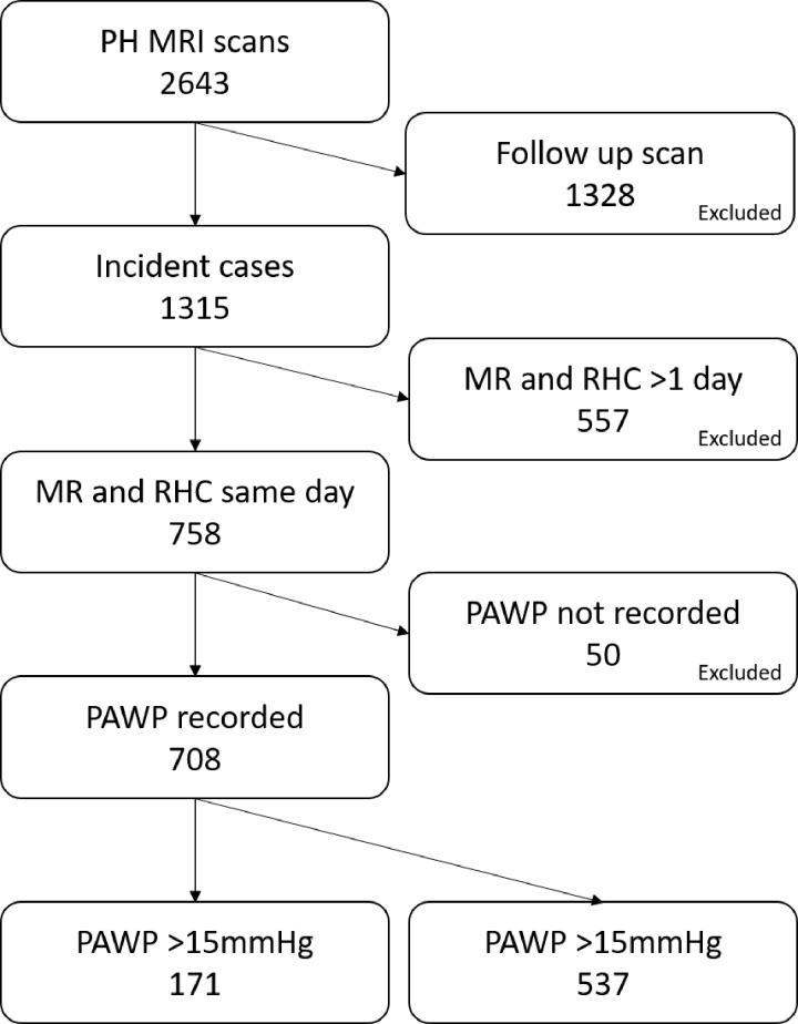
Purpose To assess interventricular septal (IVS) angle in the identification of combined pre- and postcapillary pulmonary hypertension (Cpc-PH) in patients with pulmonary hypertension (PH) due to left-sided heart disease. Materials and Methods In this retrospective study, consecutive, incident patients suspected of having PH underwent same-day right-sided heart catheterization (RHC) and MRI at a PH referral center between April 2012 and April 2017. The diagnostic accuracy of the IVS angle to identify Cpc-PH in patients with pulmonary arterial wedge pressure (PAWP) greater than 15 mmHg was assessed by using receiver operator characteristic curves, sensitivity, specificity, and negative and positive predictive values. IVS angle also was assessed as a predictor of all-cause mortality by using Cox uni- and multivariable proportional hazards regression. Results A total of 708 patients underwent same-day MRI and RHC, and 171 patients had PAWP greater than 15 mmHg. Mean age was 70 years (range, 21-90 years) (women: mean age, 69 years; range, 21-88 years) (men: mean age, 71 years; range, 43-90 years). Systolic IVS angle correlated with diastolic pulmonary gradient (DPG) (r = 0.739, P < .001). Receiver operating characteristic curve analysis showed septal angle enabled identification of Cpc-PH (DPG ≥ 7), with an area under the receiver operating characteristic curve of 0.911 (P < .001). A 160° threshold, derived from the first half of patients with raised PAWP, enabled identification of a DPG of at least 7 mmHg with 67% sensitivity and 93% specificity (P < .001) in the second cohort of patients with raised PAWP. IVS angle was predictive of all-cause mortality (standardized univariable hazard ratio, 1.615; P < .01). Conclusion The systolic interventricular septal angle is elevated in patients with combined pre- and postcapillary pulmonary hypertension and enables one to predict those patients who have PH due to left-sided heart disease who have an increased risk of death. Published under a CC BY 4.0 license. Online supplemental material is available for this article.

摘要

目的 评估室间隔角(IVS)在识别因左心疾病导致肺动脉高压(PH)患者的毛细血管前和毛细血管后肺动脉高压(Cpc-PH)中的作用。

材料与方法 在这项回顾性研究中,于 2012 年 4 月至 2017 年 4 月期间,在 PH 转诊中心,对连续出现的疑似 PH 且肺动脉楔压(PAWP)>15mmHg 的患者进行了同日右心导管检查(RHC)和 MRI。使用受试者工作特征曲线、灵敏度、特异性、阴性和阳性预测值评估 IVS 角度识别 PAWP>15mmHg 的患者中 Cpc-PH 的诊断准确性。还使用 Cox 单变量和多变量比例风险回归评估 IVS 角度作为全因死亡率的预测因子。

结果 共有 708 例患者同日进行了 MRI 和 RHC,171 例患者的 PAWP>15mmHg。平均年龄为 70 岁(范围,21-90 岁)(女性:平均年龄,69 岁;范围,21-88 岁)(男性:平均年龄,71 岁;范围,43-90 岁)。收缩期 IVS 角度与舒张性肺梯度(DPG)相关(r=0.739,P<.001)。受试者工作特征曲线分析表明,室间隔角度能够识别 Cpc-PH(DPG≥7),其受试者工作特征曲线下面积为 0.911(P<.001)。从 PAWP 升高的患者前半部分中得出的 160°阈值,可使 DPG 至少为 7mmHg,在 PAWP 升高的第二组患者中,灵敏度为 67%,特异性为 93%(P<.001)。IVS 角度可预测全因死亡率(标准化单变量危险比,1.615;P<.01)。

结论 在合并毛细血管前和毛细血管后肺动脉高压的患者中,收缩期室间隔角升高,可预测因左心疾病导致 PH 的患者中存在死亡风险增加的患者。

发表于 CC BY 4.0 许可证下。本文的在线补充材料可在网上获取。

https://cdn.ncbi.nlm.nih.gov/pmc/blobs/3c8a/6190488/99f78630db96/radiol.2018180120.fig4.jpg
https://cdn.ncbi.nlm.nih.gov/pmc/blobs/3c8a/6190488/fe8946df7872/radiol.2018180120.fig1.jpg
https://cdn.ncbi.nlm.nih.gov/pmc/blobs/3c8a/6190488/ab2489608a52/radiol.2018180120.fig2.jpg
https://cdn.ncbi.nlm.nih.gov/pmc/blobs/3c8a/6190488/f6b79323782c/radiol.2018180120.fig3.jpg
https://cdn.ncbi.nlm.nih.gov/pmc/blobs/3c8a/6190488/99f78630db96/radiol.2018180120.fig4.jpg

相似文献

1
Identifying At-Risk Patients with Combined Pre- and Postcapillary Pulmonary Hypertension Using Interventricular Septal Angle at Cardiac MRI.使用心脏 MRI 中的室间隔角识别合并毛细血管前和毛细血管后肺动脉高压的高危患者。
Radiology. 2018 Oct;289(1):61-68. doi: 10.1148/radiol.2018180120. Epub 2018 Jul 3.
2
Diagnosis of Pulmonary Hypertension with Cardiac MRI: Derivation and Validation of Regression Models.心脏 MRI 诊断肺动脉高压:回归模型的建立和验证。
Radiology. 2019 Jan;290(1):61-68. doi: 10.1148/radiol.2018180603. Epub 2018 Oct 23.
3
Pulmonary hypertension: accuracy of detection with left ventricular septal-to-free wall curvature ratio measured at cardiac MR.肺动脉高压:心脏磁共振测量左心室室间隔与游离壁曲率比检测的准确性
Radiology. 2007 Apr;243(1):63-9. doi: 10.1148/radiol.2431060067.
4
Diastolic Pressure Difference to Classify Pulmonary Hypertension in the Assessment of Heart Transplant Candidates.在评估心脏移植候选者时,用舒张压差异来分类肺动脉高压。
Circ Heart Fail. 2017 Sep;10(9). doi: 10.1161/CIRCHEARTFAILURE.117.004077.
5
A critical appraisal of transpulmonary and diastolic pressure gradients.经肺压与舒张压梯度的批判性评估。
Physiol Rep. 2016 Sep;4(17). doi: 10.14814/phy2.12910.
6
Determinants and prognostic implications of the negative diastolic pulmonary pressure gradient in patients with pulmonary hypertension due to left heart disease.左心疾病所致肺动脉高压患者中负舒张压肺压力梯度的决定因素及其预后意义。
Eur J Heart Fail. 2017 Jan;19(1):88-97. doi: 10.1002/ejhf.675. Epub 2016 Oct 17.
7
A simple echocardiographic score for the diagnosis of pulmonary vascular disease in heart failure.一种用于诊断心力衰竭中肺血管疾病的简单超声心动图评分。
J Cardiovasc Med (Hagerstown). 2017 Apr;18(4):237-243. doi: 10.2459/JCM.0000000000000485.
8
Ventilatory power, a cardiopulmonary exercise testing parameter for the prediction of pulmonary hypertension at right heart catheterization.通气功率,一种用于预测右心导管检查时肺动脉高压的心肺运动试验参数。
Int J Cardiol Heart Vasc. 2020 Apr 21;28:100513. doi: 10.1016/j.ijcha.2020.100513. eCollection 2020 Jun.
9
Three-dimensional echocardiography and 2D-3D speckle-tracking imaging in chronic pulmonary hypertension: diagnostic accuracy in detecting hemodynamic signs of right ventricular (RV) failure.三维超声心动图和二维-三维斑点追踪成像在慢性肺动脉高压中的应用:检测右心室(RV)衰竭血流动力学征象的诊断准确性
J Am Heart Assoc. 2015 Mar 19;4(3):e001584. doi: 10.1161/JAHA.114.001584.
10
Noninvasive estimation of PA pressure, flow, and resistance with CMR imaging: derivation and prospective validation study from the ASPIRE registry.应用 CMR 成像无创估计肺动脉压、流量和阻力:来自 ASPIRE 注册研究的推导和前瞻性验证。
JACC Cardiovasc Imaging. 2013 Oct;6(10):1036-1047. doi: 10.1016/j.jcmg.2013.01.013. Epub 2013 Jun 13.

引用本文的文献

1
Ventricular interdependent phenotype of mixed Cpc-pulmonary hypertension and HFpEF with normal left atrium: Impact on CPET metrics and clinical outcomes.伴有正常左心房的混合性Cpc-肺动脉高压和射血分数保留的心力衰竭的心室相互依赖表型:对心肺运动试验指标和临床结局的影响
Pulm Circ. 2024 Oct 11;14(4):e12449. doi: 10.1002/pul2.12449. eCollection 2024 Oct.
2
The Role of Cardiac MRI in Pulmonary Hypertension- Is it Still an Underutilized Tool.心脏磁共振成像在肺动脉高压中的作用——它仍然是一种未被充分利用的工具吗?
Open Respir Med J. 2024 Jun 4;18:e18743064288565. doi: 10.2174/0118743064288565240515115239. eCollection 2024.
3
Two prospective, multicenter studies for the identification of biomarker signatures for early detection of pulmonary hypertension (PH): The CIPHER and CIPHER-MRI studies.

本文引用的文献

1
Current and emerging imaging techniques in the diagnosis and assessment of pulmonary hypertension.当前和新兴的影像学技术在肺动脉高压的诊断和评估中的应用。
Expert Rev Respir Med. 2018 Feb;12(2):145-160. doi: 10.1080/17476348.2018.1420478.
2
Non-invasive methods for estimating mPAP in COPD using cardiovascular magnetic resonance imaging.使用心血管磁共振成像估计 COPD 患者中 mPAP 的无创方法。
Eur Radiol. 2018 Apr;28(4):1438-1448. doi: 10.1007/s00330-017-5143-y. Epub 2017 Nov 16.
3
Pulmonary Hypertension in Heart Failure: Pathophysiology, Pathobiology, and Emerging Clinical Perspectives.
两项用于识别肺动脉高压(PH)早期检测生物标志物特征的前瞻性多中心研究:CIPHER研究和CIPHER-MRI研究。
Pulm Circ. 2024 Jun 12;14(2):e12386. doi: 10.1002/pul2.12386. eCollection 2024 Apr.
4
Clinical-imaging-pathological correlation in pulmonary hypertension associated with left heart disease.左心疾病相关肺动脉高压的临床-影像-病理相关性。
Eur Respir Rev. 2024 Feb 28;33(171). doi: 10.1183/16000617.0144-2023. Print 2024 Jan 31.
5
Cardiac magnetic resonance assessment of cardiac involvement in autoimmune diseases.自身免疫性疾病心脏受累的心脏磁共振评估
Front Cardiovasc Med. 2023 Sep 22;10:1215907. doi: 10.3389/fcvm.2023.1215907. eCollection 2023.
6
Elucidating the Clinical Implications and Pathophysiology of Pulmonary Hypertension in Heart Failure With Preserved Ejection Fraction: A Call to Action: A Science Advisory From the American Heart Association.阐述射血分数保留的心力衰竭中肺动脉高压的临床意义和病理生理学:行动呼吁:美国心脏协会的科学声明。
Circulation. 2022 Aug 16;146(7):e73-e88. doi: 10.1161/CIR.0000000000001079. Epub 2022 Jul 11.
7
CMR Measures of Left Atrial Volume Index and Right Ventricular Function Have Prognostic Value in Chronic Thromboembolic Pulmonary Hypertension.左心房容积指数和右心室功能的心脏磁共振成像测量对慢性血栓栓塞性肺动脉高压具有预后价值。
Front Med (Lausanne). 2022 Mar 14;9:840196. doi: 10.3389/fmed.2022.840196. eCollection 2022.
8
Clinical Application of 4D Flow MR Imaging to Pulmonary Hypertension.4D 流磁共振成像在肺动脉高压中的临床应用。
Magn Reson Med Sci. 2022 Mar 1;21(2):309-318. doi: 10.2463/mrms.rev.2021-0111. Epub 2022 Feb 19.
9
Cardiac Magnetic Resonance in Pulmonary Hypertension-an Update.肺动脉高压的心脏磁共振成像——最新进展
Curr Cardiovasc Imaging Rep. 2020;13(12):30. doi: 10.1007/s12410-020-09550-2. Epub 2020 Nov 7.
10
Diagnostic accuracy of CT pulmonary angiography in suspected pulmonary hypertension.CT肺血管造影在疑似肺动脉高压中的诊断准确性
Eur Radiol. 2020 Sep;30(9):4918-4929. doi: 10.1007/s00330-020-06846-1. Epub 2020 Apr 27.
心力衰竭相关肺动脉高压:病理生理学、病理生物学与新兴临床观点。
J Am Coll Cardiol. 2017 Apr 4;69(13):1718-1734. doi: 10.1016/j.jacc.2017.01.051.
4
Magnetic Resonance Imaging in the Prognostic Evaluation of Patients with Pulmonary Arterial Hypertension.磁共振成像在肺动脉高压患者预后评估中的应用
Am J Respir Crit Care Med. 2017 Jul 15;196(2):228-239. doi: 10.1164/rccm.201611-2365OC.
5
Balancing the positives and negatives of the diastolic pulmonary gradient.权衡舒张期肺梯度的利弊。
Eur J Heart Fail. 2017 Jan;19(1):98-100. doi: 10.1002/ejhf.704.
6
Biological Phenotyping of Combined Post-Capillary and Pre-Capillary Pulmonary Hypertension: Focus on Pulmonary Vascular Remodeling.毛细血管后和毛细血管前合并性肺动脉高压的生物学表型:聚焦于肺血管重塑
J Am Coll Cardiol. 2016 Dec 13;68(23):2537-2539. doi: 10.1016/j.jacc.2016.10.013.
7
Clinical and Biological Insights Into Combined Post- and Pre-Capillary Pulmonary Hypertension.对毛细血管后和毛细血管前合并性肺动脉高压的临床与生物学见解。
J Am Coll Cardiol. 2016 Dec 13;68(23):2525-2536. doi: 10.1016/j.jacc.2016.09.942.
8
Pilot Study of Endothelin Receptor Blockade in Heart Failure with Diastolic Dysfunction and Pulmonary Hypertension (BADDHY-Trial).舒张功能障碍和肺动脉高压性心力衰竭中内皮素受体阻断的初步研究(BADDHY试验)
Heart Lung Circ. 2017 May;26(5):433-441. doi: 10.1016/j.hlc.2016.09.004. Epub 2016 Sep 28.
9
Determinants and prognostic implications of the negative diastolic pulmonary pressure gradient in patients with pulmonary hypertension due to left heart disease.左心疾病所致肺动脉高压患者中负舒张压肺压力梯度的决定因素及其预后意义。
Eur J Heart Fail. 2017 Jan;19(1):88-97. doi: 10.1002/ejhf.675. Epub 2016 Oct 17.
10
Pre-Capillary, Combined, and Post-Capillary Pulmonary Hypertension: A Pathophysiological Continuum.毛细血管前、混合性和毛细血管后肺动脉高压:一种病理生理连续统。
J Am Coll Cardiol. 2016 Jul 26;68(4):368-78. doi: 10.1016/j.jacc.2016.05.047.