Donabedian Patrick L, Kossatz Susanne, Engelbach John A, Jannetti Stephen A, Carney Brandon, Young Robert J, Weber Wolfgang A, Garbow Joel R, Reiner Thomas
Department of Radiology, Memorial Sloan-Kettering Cancer Center, 1275 York Avenue, New York, NY, 10065, USA.
Department of Radiology, Washington University, St. Louis, MO, USA.
EJNMMI Res. 2018 Jul 4;8(1):59. doi: 10.1186/s13550-018-0399-z.
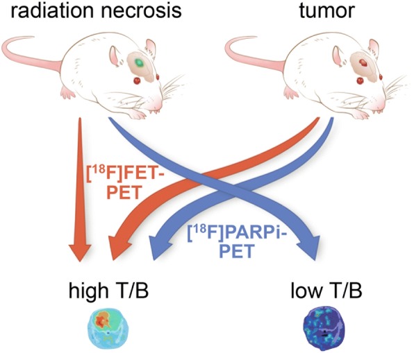
Radiation injury can be indistinguishable from recurrent tumor on standard imaging. Current protocols for this differential diagnosis require one or more follow-up imaging studies, long dynamic acquisitions, or complex image post-processing; despite much research, the inability to confidently distinguish between these two entities continues to pose a significant dilemma for the treating clinician. Using mouse models of both glioblastoma and radiation necrosis, we tested the potential of poly(ADP-ribose) polymerase (PARP)-targeted PET imaging with [F]PARPi to better discriminate radiation injury from tumor.
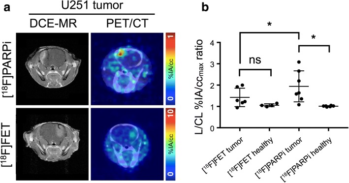
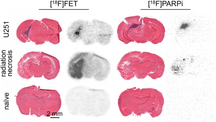
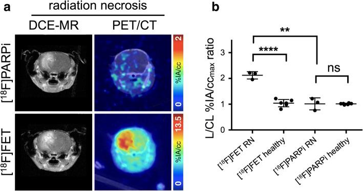
In mice with experimental radiation necrosis, lesion uptake on [F]PARPi-PET was similar to contralateral uptake (1.02 ± 0.26 lesion/contralateral %IA/cc ratio), while [F]FET-PET clearly delineated the contrast-enhancing region on MR (2.12 ± 0.16 lesion/contralateral %IA/cc ratio). In mice with focal intracranial U251 xenografts, tumor visualization on PARPi-PET was superior to FET-PET, and lesion-to-contralateral activity ratios (max/max, p = 0.034) were higher on PARPi-PET than on FET-PET.
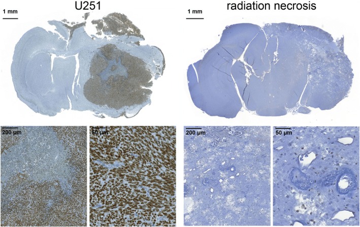
A murine model of radiation necrosis does not demonstrate [F]PARPi avidity, and [F]PARPi-PET is better than [F]FET-PET in distinguishing radiation injury from brain tumor. [F]PARPi-PET can be used for discrimination between recurrent tumor and radiation injury within a single, static imaging session, which may be of value to resolve a common dilemma in neuro-oncology.
在标准成像上,放射性损伤可能与复发性肿瘤难以区分。目前用于这种鉴别诊断的方案需要一项或多项随访成像研究、长时间动态采集或复杂的图像后处理;尽管进行了大量研究,但无法可靠地区分这两种情况仍然给临床治疗医生带来重大困境。我们使用胶质母细胞瘤和放射性坏死的小鼠模型,测试了用[F]PARPi进行聚(ADP-核糖)聚合酶(PARP)靶向PET成像以更好地区分放射性损伤和肿瘤的潜力。
在实验性放射性坏死的小鼠中,[F]PARPi-PET上病变摄取与对侧摄取相似(病变/对侧%IA/cc比值为1.02±0.26),而[F]FET-PET在磁共振成像上清晰勾勒出强化区域(病变/对侧%IA/cc比值为2.12±0.16)。在局灶性颅内U251异种移植小鼠中,PARPi-PET上的肿瘤可视化优于FET-PET,PARPi-PET上的病变与对侧活性比值(最大值/最大值,p=0.034)高于FET-PET。
放射性坏死的小鼠模型未显示[F]PARPi亲和力,[F]PARPi-PET在区分放射性损伤和脑肿瘤方面优于[F]FET-PET。[F]PARPi-PET可用于在单次静态成像过程中区分复发性肿瘤和放射性损伤,这可能有助于解决神经肿瘤学中的一个常见难题。