• 文献检索
  • 文档翻译
  • 深度研究
  • 学术资讯
  • Suppr Zotero 插件Zotero 插件
  • 邀请有礼
  • 套餐&价格
  • 历史记录
应用&插件
Suppr Zotero 插件Zotero 插件浏览器插件Mac 客户端Windows 客户端微信小程序
定价
高级版会员购买积分包购买API积分包
服务
文献检索文档翻译深度研究API 文档MCP 服务
关于我们
关于 Suppr公司介绍联系我们用户协议隐私条款
关注我们

Suppr 超能文献

核心技术专利:CN118964589B侵权必究
粤ICP备2023148730 号-1Suppr @ 2026

文献检索

告别复杂PubMed语法,用中文像聊天一样搜索,搜遍4000万医学文献。AI智能推荐,让科研检索更轻松。

立即免费搜索

文件翻译

保留排版,准确专业,支持PDF/Word/PPT等文件格式,支持 12+语言互译。

免费翻译文档

深度研究

AI帮你快速写综述,25分钟生成高质量综述,智能提取关键信息,辅助科研写作。

立即免费体验

全面分析肺癌病理图像,发现预测生存结果的肿瘤形状和边界特征。

Comprehensive analysis of lung cancer pathology images to discover tumor shape and boundary features that predict survival outcome.

机构信息

Quantitative Biomedical Research Center, Department of Clinical Sciences, University of Texas Southwestern Medical Center, Dallas, Texas, 75390, USA.

Department of Computer Sciences, Massachusetts Institute of Technology, Cambridge, MA, USA.

出版信息

Sci Rep. 2018 Jul 10;8(1):10393. doi: 10.1038/s41598-018-27707-4.

DOI:10.1038/s41598-018-27707-4
PMID:29991684
原文链接:https://pmc.ncbi.nlm.nih.gov/articles/PMC6039531/
Abstract

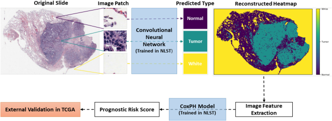
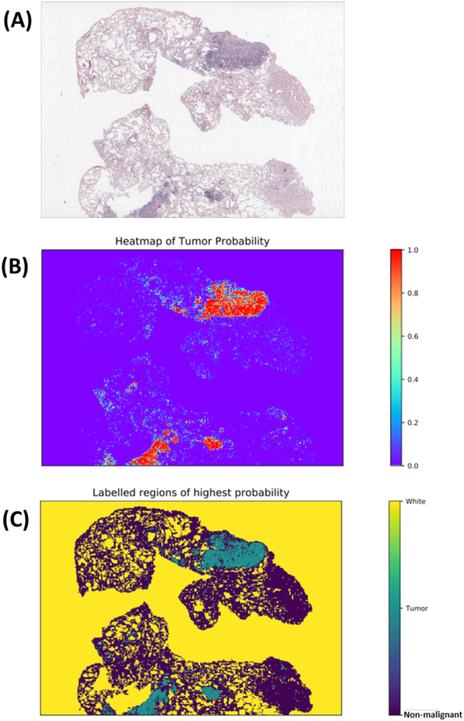
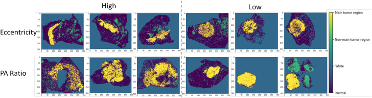
Pathology images capture tumor histomorphological details in high resolution. However, manual detection and characterization of tumor regions in pathology images is labor intensive and subjective. Using a deep convolutional neural network (CNN), we developed an automated tumor region recognition system for lung cancer pathology images. From the identified tumor regions, we extracted 22 well-defined shape and boundary features and found that 15 of them were significantly associated with patient survival outcome in lung adenocarcinoma patients from the National Lung Screening Trial. A tumor region shape-based prognostic model was developed and validated in an independent patient cohort (n = 389). The predicted high-risk group had significantly worse survival than the low-risk group (p value = 0.0029). Predicted risk group serves as an independent prognostic factor (high-risk vs. low-risk, hazard ratio = 2.25, 95% CI 1.34-3.77, p value = 0.0022) after adjusting for age, gender, smoking status, and stage. This study provides new insights into the relationship between tumor shape and patient prognosis.

摘要

病理学图像以高分辨率捕捉肿瘤组织形态学细节。然而,手动检测和描绘病理学图像中的肿瘤区域既费力又主观。我们使用深度卷积神经网络(CNN)开发了一种用于肺癌病理学图像的自动肿瘤区域识别系统。从识别的肿瘤区域中,我们提取了 22 个定义明确的形状和边界特征,发现在来自国家肺癌筛查试验的肺腺癌患者中,其中 15 个特征与患者生存结果显著相关。在一个独立的患者队列(n=389)中开发并验证了基于肿瘤区域形状的预后模型。预测的高危组的生存明显差于低危组(p 值=0.0029)。预测的风险组在调整年龄、性别、吸烟状况和分期后是一个独立的预后因素(高危 vs. 低危,风险比=2.25,95%CI 1.34-3.77,p 值=0.0022)。本研究为肿瘤形状与患者预后之间的关系提供了新的见解。

https://cdn.ncbi.nlm.nih.gov/pmc/blobs/33c8/6039531/75843aede034/41598_2018_27707_Fig4_HTML.jpg
https://cdn.ncbi.nlm.nih.gov/pmc/blobs/33c8/6039531/b353b53f8732/41598_2018_27707_Fig1_HTML.jpg
https://cdn.ncbi.nlm.nih.gov/pmc/blobs/33c8/6039531/a1cb1bb56295/41598_2018_27707_Fig2_HTML.jpg
https://cdn.ncbi.nlm.nih.gov/pmc/blobs/33c8/6039531/0bbc997f137f/41598_2018_27707_Fig3_HTML.jpg
https://cdn.ncbi.nlm.nih.gov/pmc/blobs/33c8/6039531/75843aede034/41598_2018_27707_Fig4_HTML.jpg

相似文献

1
Comprehensive analysis of lung cancer pathology images to discover tumor shape and boundary features that predict survival outcome.全面分析肺癌病理图像,发现预测生存结果的肿瘤形状和边界特征。
Sci Rep. 2018 Jul 10;8(1):10393. doi: 10.1038/s41598-018-27707-4.
2
ConvPath: A software tool for lung adenocarcinoma digital pathological image analysis aided by a convolutional neural network.ConvPath:一种使用卷积神经网络辅助肺腺癌数字病理图像分析的软件工具。
EBioMedicine. 2019 Dec;50:103-110. doi: 10.1016/j.ebiom.2019.10.033. Epub 2019 Nov 22.
3
Development and Validation of a Pathology Image Analysis-based Predictive Model for Lung Adenocarcinoma Prognosis - A Multi-cohort Study.基于病理图像分析的肺腺癌预后预测模型的建立与验证——一项多队列研究。
Sci Rep. 2019 May 3;9(1):6886. doi: 10.1038/s41598-019-42845-z.
4
Comprehensive Computational Pathological Image Analysis Predicts Lung Cancer Prognosis.综合计算病理图像分析可预测肺癌预后。
J Thorac Oncol. 2017 Mar;12(3):501-509. doi: 10.1016/j.jtho.2016.10.017. Epub 2016 Nov 4.
5
Predictors of Pathologic Tumor Invasion and Prognosis for Ground Glass Opacity Featured Lung Adenocarcinoma.预测表现为磨玻璃密度的肺腺癌的病理性肿瘤侵袭和预后的因素。
Ann Thorac Surg. 2018 Dec;106(6):1682-1690. doi: 10.1016/j.athoracsur.2018.06.058. Epub 2018 Aug 8.
6
Comparing clinicopathological features and prognosis of primary pulmonary invasive mucinous adenocarcinoma based on computed tomography findings.基于 CT 表现比较原发性肺浸润性黏液腺癌的临床病理特征和预后。
Cancer Imaging. 2019 Jul 10;19(1):47. doi: 10.1186/s40644-019-0236-2.
7
Tumor spread through air spaces is a useful predictor of recurrence and prognosis in stage I lung squamous cell carcinoma, but not in stage II and III.肿瘤通过气腔播散是 I 期肺鳞癌复发和预后的有用预测指标,但在 II 期和 III 期则不然。
Lung Cancer. 2018 Jun;120:14-21. doi: 10.1016/j.lungcan.2018.03.018. Epub 2018 Mar 20.
8
Prognostic Value of Thyroid Transcription Factor-1 Expression in Patients with Advanced Lung Adenocarcinoma.甲状腺转录因子-1表达在晚期肺腺癌患者中的预后价值
In Vivo. 2018 Nov-Dec;32(6):1571-1579. doi: 10.21873/invivo.11416.
9
Classification of lung adenocarcinoma transcriptome subtypes from pathological images using deep convolutional networks.利用深度卷积网络从病理图像中对肺腺癌转录组亚型进行分类。
Int J Comput Assist Radiol Surg. 2018 Dec;13(12):1905-1913. doi: 10.1007/s11548-018-1835-2. Epub 2018 Aug 29.
10
Central Tumor Location at Chest CT Is an Adverse Prognostic Factor for Disease-Free Survival of Node-Negative Early-Stage Lung Adenocarcinomas.胸部 CT 中央肿瘤位置是淋巴结阴性早期肺腺癌无病生存的不良预后因素。
Radiology. 2021 May;299(2):438-447. doi: 10.1148/radiol.2021203937. Epub 2021 Feb 23.

引用本文的文献

1
Radiomic Features Prognosticate Treatment Response in CAR-T Cell Therapy.放射组学特征可预测CAR-T细胞疗法的治疗反应。
Cancers (Basel). 2025 May 30;17(11):1832. doi: 10.3390/cancers17111832.
2
A promptable CT foundation model for solid tumor evaluation.一种用于实体瘤评估的可提示式CT基础模型。
NPJ Precis Oncol. 2025 Apr 25;9(1):121. doi: 10.1038/s41698-025-00903-y.
3
Identifying invasiveness to aid lung adenocarcinoma diagnosis using deep learning and pathomics.利用深度学习和病理组学识别侵袭性以辅助肺腺癌诊断

本文引用的文献

1
Deep learning in breast cancer risk assessment: evaluation of convolutional neural networks on a clinical dataset of full-field digital mammograms.深度学习在乳腺癌风险评估中的应用:基于全场数字化乳腺X线摄影临床数据集对卷积神经网络的评估
J Med Imaging (Bellingham). 2017 Oct;4(4):041304. doi: 10.1117/1.JMI.4.4.041304. Epub 2017 Sep 13.
2
Accurate and reproducible invasive breast cancer detection in whole-slide images: A Deep Learning approach for quantifying tumor extent.全切片图像中精确且可重复的浸润性乳腺癌检测:一种用于量化肿瘤范围的深度学习方法。
Sci Rep. 2017 Apr 18;7:46450. doi: 10.1038/srep46450.
3
Network Analysis Reveals A Signaling Regulatory Loop in the PIK3CA-mutated Breast Cancer Predicting Survival Outcome.
Sci Rep. 2025 Feb 10;15(1):4913. doi: 10.1038/s41598-025-87094-5.
4
Deep learning analysis of histopathological images predicts immunotherapy prognosis and reveals tumour microenvironment features in non-small cell lung cancer.深度学习分析组织病理学图像预测非小细胞肺癌的免疫治疗预后并揭示肿瘤微环境特征。
Br J Cancer. 2024 Dec;131(11):1833-1845. doi: 10.1038/s41416-024-02856-8. Epub 2024 Oct 25.
5
Bayesian Landmark-based Shape Analysis of Tumor Pathology Images.基于贝叶斯地标点的肿瘤病理图像形状分析
J Am Stat Assoc. 2024;119(546):798-810. doi: 10.1080/01621459.2023.2298031. Epub 2024 Feb 1.
6
Antibody-Mediated Rejection in Lung Transplantation: Diagnosis and Therapeutic Armamentarium in a 21st Century Perspective.抗体介导的肺移植排斥反应:21 世纪视角下的诊断和治疗策略。
Transpl Int. 2024 Aug 7;37:12973. doi: 10.3389/ti.2024.12973. eCollection 2024.
7
A Pan-Cancer Patient-Derived Xenograft Histology Image Repository with Genomic and Pathologic Annotations Enables Deep Learning Analysis.Pan-Cancer 患者来源异种移植组织病理学图像库与基因组和病理学注释相结合,可实现深度学习分析。
Cancer Res. 2024 Jul 2;84(13):2060-2072. doi: 10.1158/0008-5472.CAN-23-1349.
8
Unveiling the landscape of pathomics in personalized immunotherapy for lung cancer: a bibliometric analysis.揭示肺癌个性化免疫治疗中病理组学的全景:一项文献计量分析。
Front Oncol. 2024 Jul 8;14:1432212. doi: 10.3389/fonc.2024.1432212. eCollection 2024.
9
Mapping the landscape of histomorphological cancer phenotypes using self-supervised learning on unannotated pathology slides.利用无标注病理切片的自监督学习来绘制癌症表型的组织形态学图谱。
Nat Commun. 2024 Jun 11;15(1):4596. doi: 10.1038/s41467-024-48666-7.
10
Implementation of Artificial Intelligence in Personalized Prognostic Assessment of Lung Cancer: A Narrative Review.人工智能在肺癌个性化预后评估中的应用:一项叙述性综述
Cancers (Basel). 2024 May 10;16(10):1832. doi: 10.3390/cancers16101832.
网络分析揭示了PIK3CA突变型乳腺癌中预测生存结果的信号调节回路。
Genomics Proteomics Bioinformatics. 2017 Apr;15(2):121-129. doi: 10.1016/j.gpb.2017.02.002. Epub 2017 Apr 7.
4
Relevance of deep learning to facilitate the diagnosis of HER2 status in breast cancer.深度学习在乳腺癌 HER2 状态诊断中的相关性。
Sci Rep. 2017 Apr 5;7:45938. doi: 10.1038/srep45938.
5
Dermatologist-level classification of skin cancer with deep neural networks.基于深度神经网络的皮肤癌皮肤科医生级分类。
Nature. 2017 Feb 2;542(7639):115-118. doi: 10.1038/nature21056. Epub 2017 Jan 25.
6
Comprehensive Computational Pathological Image Analysis Predicts Lung Cancer Prognosis.综合计算病理图像分析可预测肺癌预后。
J Thorac Oncol. 2017 Mar;12(3):501-509. doi: 10.1016/j.jtho.2016.10.017. Epub 2016 Nov 4.
7
Predicting non-small cell lung cancer prognosis by fully automated microscopic pathology image features.通过全自动显微镜病理图像特征预测非小细胞肺癌预后。
Nat Commun. 2016 Aug 16;7:12474. doi: 10.1038/ncomms12474.
8
Regularization Paths for Cox's Proportional Hazards Model via Coordinate Descent.通过坐标下降法求解Cox比例风险模型的正则化路径
J Stat Softw. 2011 Mar;39(5):1-13. doi: 10.18637/jss.v039.i05.
9
The IASLC Lung Cancer Staging Project: Proposals for Revision of the TNM Stage Groupings in the Forthcoming (Eighth) Edition of the TNM Classification for Lung Cancer.IASLC 肺癌分期项目:对即将发布的(第八版)肺癌 TNM 分类中 TNM 分期分组的修订建议。
J Thorac Oncol. 2016 Jan;11(1):39-51. doi: 10.1016/j.jtho.2015.09.009.
10
Identification and Construction of Combinatory Cancer Hallmark-Based Gene Signature Sets to Predict Recurrence and Chemotherapy Benefit in Stage II Colorectal Cancer.基于组合癌症特征的基因签名集的鉴定和构建,以预测 II 期结直肠癌的复发和化疗获益。
JAMA Oncol. 2016 Jan;2(1):37-45. doi: 10.1001/jamaoncol.2015.3413.