Quantitative Biomedical Research Center, Department of Population and Data Sciences, University of Texas Southwestern Medical Center, Dallas, TX.
Quantitative Biomedical Research Center, Department of Population and Data Sciences, University of Texas Southwestern Medical Center, Dallas, TX; Center for the Genetics of Host Defense, University of Texas Southwestern Medical Center, Dallas, TX.
EBioMedicine. 2019 Dec;50:103-110. doi: 10.1016/j.ebiom.2019.10.033. Epub 2019 Nov 22.
The spatial distributions of different types of cells could reveal a cancer cell's growth pattern, its relationships with the tumor microenvironment and the immune response of the body, all of which represent key "hallmarks of cancer". However, the process by which pathologists manually recognize and localize all the cells in pathology slides is extremely labor intensive and error prone.
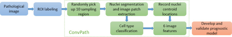
In this study, we developed an automated cell type classification pipeline, ConvPath, which includes nuclei segmentation, convolutional neural network-based tumor cell, stromal cell, and lymphocyte classification, and extraction of tumor microenvironment-related features for lung cancer pathology images. To facilitate users in leveraging this pipeline for their research, all source scripts for ConvPath software are available at https://qbrc.swmed.edu/projects/cnn/.
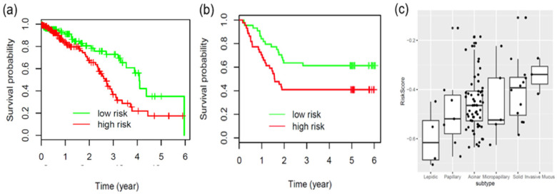
The overall classification accuracy was 92.9% and 90.1% in training and independent testing datasets, respectively. By identifying cells and classifying cell types, this pipeline can convert a pathology image into a "spatial map" of tumor, stromal and lymphocyte cells. From this spatial map, we can extract features that characterize the tumor micro-environment. Based on these features, we developed an image feature-based prognostic model and validated the model in two independent cohorts. The predicted risk group serves as an independent prognostic factor, after adjusting for clinical variables that include age, gender, smoking status, and stage.
The analysis pipeline developed in this study could convert the pathology image into a "spatial map" of tumor cells, stromal cells and lymphocytes. This could greatly facilitate and empower comprehensive analysis of the spatial organization of cells, as well as their roles in tumor progression and metastasis.
不同类型细胞的空间分布可以揭示癌细胞的生长模式、其与肿瘤微环境的关系以及机体的免疫反应,这些都是癌症的关键“特征”。然而,病理学家手动识别和定位病理载玻片上所有细胞的过程极其耗费人力且容易出错。
在这项研究中,我们开发了一种自动化细胞类型分类流水线 ConvPath,它包括细胞核分割、基于卷积神经网络的肿瘤细胞、基质细胞和淋巴细胞分类,以及提取肺癌病理图像中的肿瘤微环境相关特征。为了方便用户在研究中利用这个流水线,ConvPath 软件的所有原始脚本都可以在 https://qbrc.swmed.edu/projects/cnn/ 上获取。
在训练集和独立测试集中,整体分类准确率分别为 92.9%和 90.1%。通过识别细胞和分类细胞类型,该流水线可以将病理图像转换为肿瘤、基质和淋巴细胞细胞的“空间图谱”。从这个空间图谱中,我们可以提取出表征肿瘤微环境的特征。基于这些特征,我们开发了一种基于图像特征的预后模型,并在两个独立队列中验证了该模型。预测的风险组是独立的预后因素,可在调整包括年龄、性别、吸烟状况和分期在内的临床变量后使用。
本研究中开发的分析流水线可以将病理图像转换为肿瘤细胞、基质细胞和淋巴细胞的“空间图谱”。这将极大地促进和增强对细胞空间组织及其在肿瘤进展和转移中的作用的全面分析。