• 文献检索
  • 文档翻译
  • 深度研究
  • 学术资讯
  • Suppr Zotero 插件Zotero 插件
  • 邀请有礼
  • 套餐&价格
  • 历史记录
应用&插件
Suppr Zotero 插件Zotero 插件浏览器插件Mac 客户端Windows 客户端微信小程序
定价
高级版会员购买积分包购买API积分包
服务
文献检索文档翻译深度研究API 文档MCP 服务
关于我们
关于 Suppr公司介绍联系我们用户协议隐私条款
关注我们

Suppr 超能文献

核心技术专利:CN118964589B侵权必究
粤ICP备2023148730 号-1Suppr @ 2026

文献检索

告别复杂PubMed语法,用中文像聊天一样搜索,搜遍4000万医学文献。AI智能推荐,让科研检索更轻松。

立即免费搜索

文件翻译

保留排版,准确专业,支持PDF/Word/PPT等文件格式,支持 12+语言互译。

免费翻译文档

深度研究

AI帮你快速写综述,25分钟生成高质量综述,智能提取关键信息,辅助科研写作。

立即免费体验

内皮细胞凋亡有助于斑马鱼发育过程中的脑血管修剪。

Apoptosis of Endothelial Cells Contributes to Brain Vessel Pruning of Zebrafish During Development.

作者信息

Zhang Yu, Xu Bing, Chen Qi, Yan Yong, Du Jiulin, Du Xufei

机构信息

Institute of Neuroscience, State Key Laboratory of Neuroscience, Center for Excellence in Brain Science and Intelligence Technology, Chinese Academy of Sciences, Shanghai, China.

School of Future Technology, University of Chinese Academy of Sciences, Beijing, China.

出版信息

Front Mol Neurosci. 2018 Jun 28;11:222. doi: 10.3389/fnmol.2018.00222. eCollection 2018.

DOI:10.3389/fnmol.2018.00222
PMID:30002618
原文链接:https://pmc.ncbi.nlm.nih.gov/articles/PMC6031709/
Abstract

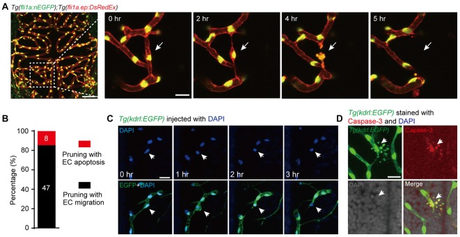
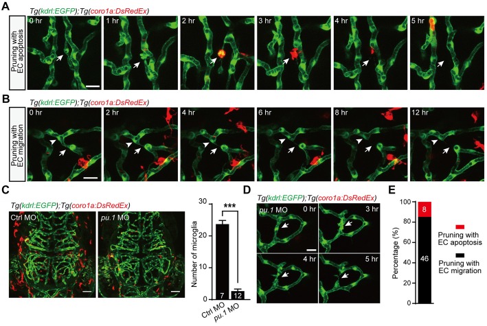
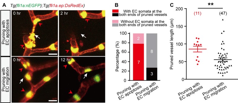
During development, immature blood vessel networks remodel to form a simplified and efficient vasculature to meet the demand for oxygen and nutrients, and this remodeling process is mainly achieved via the pruning of existing vessels. It has already known that the migration of vascular endothelial cells (ECs) is one of the mechanisms underlying vessel pruning. However, the role of EC apoptosis in vessel pruning remains under debate, especially in the brain. Here, we reported that EC apoptosis makes a significant contribution to vessel pruning in the brain of larval zebrafish. Using long-term time-lapse confocal imaging of the brain vasculature in zebrafish larvae, we found that EC apoptosis was always accompanied with brain vessel pruning and about 15% of vessel pruning events were resulted from EC apoptosis. In comparison with brain vessels undergoing EC migration-associated pruning, EC apoptosis-accompanied pruned vessels were longer and showed higher probability that the nuclei of neighboring vessels' ECs occupied their both ends. Furthermore, we found that microglia were responsible for the clearance of apoptotic ECs accompanying vessel pruning, though microglia themselves were dispensable for the occurrence of vessel pruning. Thus, our study demonstrates that EC apoptosis contributes to vessel pruning in the brain during development in a microglial cell-independent manner.

摘要

在发育过程中,不成熟的血管网络会进行重塑,以形成一个简化且高效的脉管系统,以满足对氧气和营养物质的需求,而这种重塑过程主要是通过对现有血管的修剪来实现的。已知血管内皮细胞(ECs)的迁移是血管修剪的潜在机制之一。然而,EC凋亡在血管修剪中的作用仍存在争议,尤其是在大脑中。在此,我们报告EC凋亡对斑马鱼幼体大脑中的血管修剪有显著贡献。通过对斑马鱼幼体脑血管进行长期延时共聚焦成像,我们发现EC凋亡总是伴随着脑血管修剪,并且约15%的血管修剪事件是由EC凋亡导致的。与经历与EC迁移相关修剪的脑血管相比,伴随EC凋亡而修剪的血管更长,并且相邻血管EC的细胞核占据其两端的概率更高。此外,我们发现小胶质细胞负责清除伴随血管修剪的凋亡EC,尽管小胶质细胞本身对于血管修剪的发生并非必需。因此,我们的研究表明,EC凋亡以一种不依赖小胶质细胞的方式,在发育过程中对大脑中的血管修剪有贡献。

https://cdn.ncbi.nlm.nih.gov/pmc/blobs/1014/6031709/b3fba21270b3/fnmol-11-00222-g0003.jpg
https://cdn.ncbi.nlm.nih.gov/pmc/blobs/1014/6031709/ad3fc022b354/fnmol-11-00222-g0001.jpg
https://cdn.ncbi.nlm.nih.gov/pmc/blobs/1014/6031709/c4d1644f9854/fnmol-11-00222-g0002.jpg
https://cdn.ncbi.nlm.nih.gov/pmc/blobs/1014/6031709/b3fba21270b3/fnmol-11-00222-g0003.jpg
https://cdn.ncbi.nlm.nih.gov/pmc/blobs/1014/6031709/ad3fc022b354/fnmol-11-00222-g0001.jpg
https://cdn.ncbi.nlm.nih.gov/pmc/blobs/1014/6031709/c4d1644f9854/fnmol-11-00222-g0002.jpg
https://cdn.ncbi.nlm.nih.gov/pmc/blobs/1014/6031709/b3fba21270b3/fnmol-11-00222-g0003.jpg

相似文献

1
Apoptosis of Endothelial Cells Contributes to Brain Vessel Pruning of Zebrafish During Development.内皮细胞凋亡有助于斑马鱼发育过程中的脑血管修剪。
Front Mol Neurosci. 2018 Jun 28;11:222. doi: 10.3389/fnmol.2018.00222. eCollection 2018.
2
Haemodynamics-driven developmental pruning of brain vasculature in zebrafish.血流动力学驱动斑马鱼脑血管发育性修剪。
PLoS Biol. 2012;10(8):e1001374. doi: 10.1371/journal.pbio.1001374. Epub 2012 Aug 14.
3
The role of blood flow in vessel remodeling and its regulatory mechanism during developmental angiogenesis.血流在血管重构中的作用及其在发育性血管生成中的调控机制。
Cell Mol Life Sci. 2023 May 23;80(6):162. doi: 10.1007/s00018-023-04801-z.
4
Endothelial cell self-fusion during vascular pruning.血管修剪过程中的内皮细胞自我融合。
PLoS Biol. 2015 Apr 17;13(4):e1002126. doi: 10.1371/journal.pbio.1002126. eCollection 2015 Apr.
5
The blood flow-klf6a-tagln2 axis drives vessel pruning in zebrafish by regulating endothelial cell rearrangement and actin cytoskeleton dynamics.血流-Klf6a-Tagln2 轴通过调节内皮细胞重排和肌动蛋白细胞骨架动力学来驱动斑马鱼血管修剪。
PLoS Genet. 2021 Jul 28;17(7):e1009690. doi: 10.1371/journal.pgen.1009690. eCollection 2021 Jul.
6
TMEM215 Prevents Endothelial Cell Apoptosis in Vessel Regression by Blunting BIK-Regulated ER-to-Mitochondrial Ca Influx.跨膜蛋白215通过减弱BIK调节的内质网到线粒体的钙内流来防止血管消退过程中的内皮细胞凋亡。
Circ Res. 2023 Oct 13;133(9):739-757. doi: 10.1161/CIRCRESAHA.123.322686. Epub 2023 Sep 26.
7
Apoptosis regulates endothelial cell number and capillary vessel diameter but not vessel regression during retinal angiogenesis.细胞凋亡调节视网膜血管生成过程中的内皮细胞数量和毛细血管直径,但不调节血管消退。
Development. 2016 Aug 15;143(16):2973-82. doi: 10.1242/dev.137513. Epub 2016 Jul 28.
8
Visualizing the cell-cycle progression of endothelial cells in zebrafish.观察斑马鱼内皮细胞的细胞周期进程。
Dev Biol. 2014 Sep 1;393(1):10-23. doi: 10.1016/j.ydbio.2014.06.015. Epub 2014 Jun 26.
9
Blood flow changes coincide with cellular rearrangements during blood vessel pruning in zebrafish embryos.血流变化与斑马鱼胚胎血管修剪过程中的细胞重排相吻合。
PLoS One. 2013 Oct 11;8(10):e75060. doi: 10.1371/journal.pone.0075060. eCollection 2013.
10
Endothelial cell-derived non-canonical Wnt ligands control vascular pruning in angiogenesis.内皮细胞衍生的非经典 Wnt 配体控制血管生成中的血管修剪。
Development. 2014 Apr;141(8):1757-66. doi: 10.1242/dev.104422.

引用本文的文献

1
Direct blood flow velocity measurements of zebrafish cerebral vessels with a dual-mode light-sheet microscope.使用双模式光片显微镜对斑马鱼脑血管进行直接血流速度测量。
Biomed Opt Express. 2025 Jun 2;16(7):2573-2583. doi: 10.1364/BOE.553584. eCollection 2025 Jul 1.
2
Neural Regulation of Vascular Development: Molecular Mechanisms and Interactions.神经对血管发育的调节:分子机制与相互作用。
Biomolecules. 2024 Aug 8;14(8):966. doi: 10.3390/biom14080966.
3
Social deficits mirror delayed cerebrovascular dysfunction after traumatic brain injury.

本文引用的文献

1
Cell behaviors and dynamics during angiogenesis.血管生成过程中的细胞行为与动态变化。
Development. 2016 Jul 1;143(13):2249-60. doi: 10.1242/dev.135616.
2
Macrophages Mediate the Repair of Brain Vascular Rupture through Direct Physical Adhesion and Mechanical Traction.巨噬细胞通过直接物理黏附和机械牵拉介导脑血管破裂修复。
Immunity. 2016 May 17;44(5):1162-76. doi: 10.1016/j.immuni.2016.03.008. Epub 2016 May 3.
3
Purinergic receptor P2RY12-dependent microglial closure of the injured blood-brain barrier.嘌呤能受体P2RY12依赖性小胶质细胞对受损血脑屏障的封闭作用。
社会缺陷反映了创伤性脑损伤后的脑血管功能延迟障碍。
Acta Neuropathol Commun. 2024 Aug 7;12(1):126. doi: 10.1186/s40478-024-01840-w.
4
Cellular and molecular mechanisms in vascular repair after traumatic brain injury: a narrative review.创伤性脑损伤后血管修复的细胞和分子机制:一篇叙述性综述
Burns Trauma. 2023 Sep 4;11:tkad033. doi: 10.1093/burnst/tkad033. eCollection 2023.
5
The role of blood flow in vessel remodeling and its regulatory mechanism during developmental angiogenesis.血流在血管重构中的作用及其在发育性血管生成中的调控机制。
Cell Mol Life Sci. 2023 May 23;80(6):162. doi: 10.1007/s00018-023-04801-z.
6
Physiological and Pathological Remodeling of Cerebral Microvessels.脑微血管的生理和病理重塑。
Int J Mol Sci. 2022 Oct 21;23(20):12683. doi: 10.3390/ijms232012683.
7
Identification of Potential Core Genes for the Rupture of Intracranial Aneurysms by a Bioinformatics Analysis.通过生物信息学分析鉴定颅内动脉瘤破裂的潜在核心基因
Front Genet. 2022 Mar 30;13:875007. doi: 10.3389/fgene.2022.875007. eCollection 2022.
8
Anatomy and development of the pectoral fin vascular network in the zebrafish.斑马鱼胸鳍血管网络的解剖结构和发育。
Development. 2022 Mar 1;149(5). doi: 10.1242/dev.199676. Epub 2022 Mar 4.
9
Effects of Di-Isononyl Phthalate (DiNP) on Follicular Atresia in Zebrafish Ovary.邻苯二甲酸二异壬酯(DiNP)对斑马鱼卵巢滤泡闭锁的影响。
Front Endocrinol (Lausanne). 2021 Jun 14;12:677853. doi: 10.3389/fendo.2021.677853. eCollection 2021.
10
Contribution of cell death signaling to blood vessel formation.细胞死亡信号通路对血管生成的贡献。
Cell Mol Life Sci. 2021 Apr;78(7):3247-3264. doi: 10.1007/s00018-020-03738-x. Epub 2021 Mar 30.
Proc Natl Acad Sci U S A. 2016 Jan 26;113(4):1074-9. doi: 10.1073/pnas.1520398113. Epub 2016 Jan 11.
4
Microglia: Dynamic Mediators of Synapse Development and Plasticity.小胶质细胞:突触发育和可塑性的动态调节因子
Trends Immunol. 2015 Oct;36(10):605-613. doi: 10.1016/j.it.2015.08.008.
5
Mechanisms of Vessel Pruning and Regression.血管修剪和退化的机制。
Dev Cell. 2015 Jul 6;34(1):5-17. doi: 10.1016/j.devcel.2015.06.004.
6
Endothelial cell self-fusion during vascular pruning.血管修剪过程中的内皮细胞自我融合。
PLoS Biol. 2015 Apr 17;13(4):e1002126. doi: 10.1371/journal.pbio.1002126. eCollection 2015 Apr.
7
Dynamic endothelial cell rearrangements drive developmental vessel regression.动态内皮细胞重排驱动发育性血管消退。
PLoS Biol. 2015 Apr 17;13(4):e1002125. doi: 10.1371/journal.pbio.1002125. eCollection 2015 Apr.
8
Microglia development and function.小胶质细胞的发育与功能。
Annu Rev Immunol. 2014;32:367-402. doi: 10.1146/annurev-immunol-032713-120240. Epub 2014 Jan 22.
9
Blood flow changes coincide with cellular rearrangements during blood vessel pruning in zebrafish embryos.血流变化与斑马鱼胚胎血管修剪过程中的细胞重排相吻合。
PLoS One. 2013 Oct 11;8(10):e75060. doi: 10.1371/journal.pone.0075060. eCollection 2013.
10
The importance of microglia in the development of the vasculature in the central nervous system.小胶质细胞在中枢神经系统血管形成中的重要性。
Vasc Cell. 2013 Feb 19;5(1):4. doi: 10.1186/2045-824X-5-4.