• 文献检索
  • 文档翻译
  • 深度研究
  • 学术资讯
  • Suppr Zotero 插件Zotero 插件
  • 邀请有礼
  • 套餐&价格
  • 历史记录
应用&插件
Suppr Zotero 插件Zotero 插件浏览器插件Mac 客户端Windows 客户端微信小程序
定价
高级版会员购买积分包购买API积分包
服务
文献检索文档翻译深度研究API 文档MCP 服务
关于我们
关于 Suppr公司介绍联系我们用户协议隐私条款
关注我们

Suppr 超能文献

核心技术专利:CN118964589B侵权必究
粤ICP备2023148730 号-1Suppr @ 2026

文献检索

告别复杂PubMed语法,用中文像聊天一样搜索,搜遍4000万医学文献。AI智能推荐,让科研检索更轻松。

立即免费搜索

文件翻译

保留排版,准确专业,支持PDF/Word/PPT等文件格式,支持 12+语言互译。

免费翻译文档

深度研究

AI帮你快速写综述,25分钟生成高质量综述,智能提取关键信息,辅助科研写作。

立即免费体验

利用活体成像研究小鼠角膜创伤评估血管生成和血管重构的细胞动力学

Characterization of multi-cellular dynamics of angiogenesis and vascular remodelling by intravital imaging of the wounded mouse cornea.

机构信息

Karolinska Institutet, Department of Medical Biochemistry and Biophysics, Division of Vascular Biology, Solnavägen 9, SE171 65, Stockholm, Sweden.

Uppsala University, Dept. Immunology, Genetics and Pathology, Rudbeck Laboratory, Dag Hammarskjölds, väg 20, SE751 85, Uppsala, Sweden.

出版信息

Sci Rep. 2018 Jul 13;8(1):10672. doi: 10.1038/s41598-018-28770-7.

DOI:10.1038/s41598-018-28770-7
PMID:30006556
原文链接:https://pmc.ncbi.nlm.nih.gov/articles/PMC6045577/
Abstract

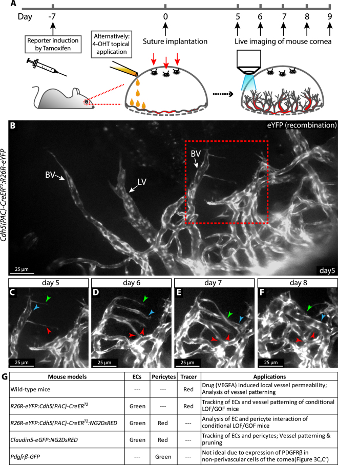
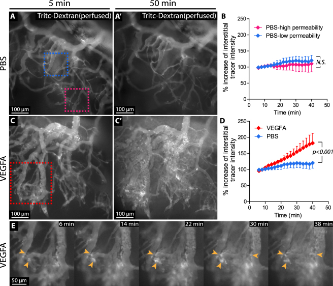
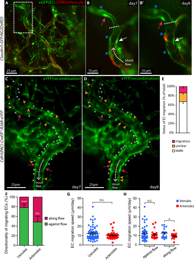
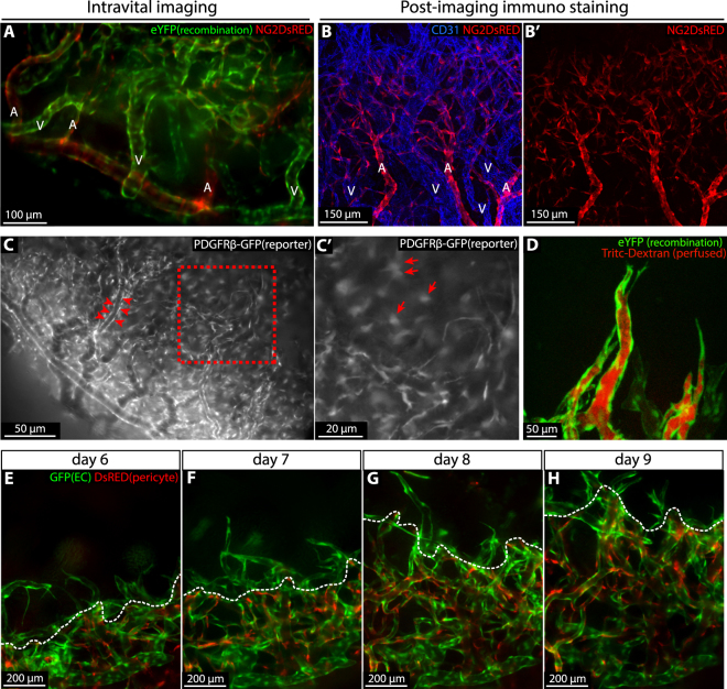
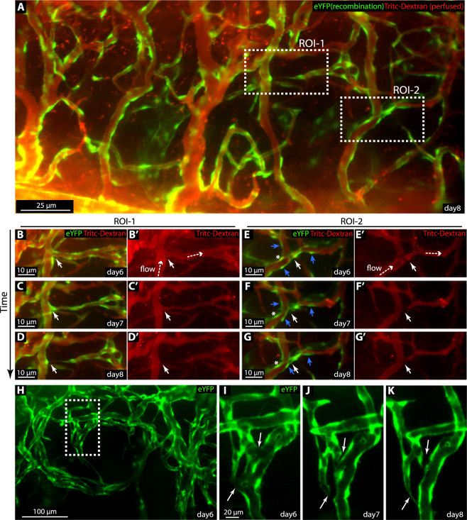
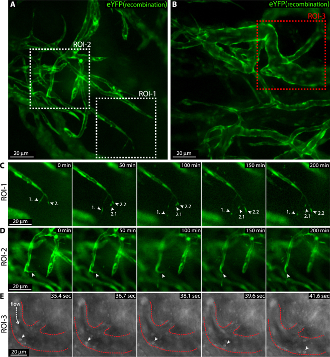
Establishment of the functional blood vasculature involves extensive cellular rearrangement controlled by growth factors, chemokines and flow-mediated shear forces. To record these highly dynamic processes in mammalians has been technically demanding. Here we apply confocal and wide field time-lapse in vivo microscopy to characterize the remodelling vasculature of the wounded mouse cornea. Using mouse lines with constitutive or inducible endogenous fluorescent reporters, in combination with tracer injections and mosaic genetic recombination, we follow processes of sprouting angiogenesis, sprout fusion, vessel expansion and pruning in vivo, at subcellular resolution. We describe the migratory behaviour of endothelial cells of perfused vessels, in relation to blood flow directionality and vessel identity. Live-imaging following intravascular injection of fluorescent tracers, allowed for recording of VEGFA-induced permeability. Altogether, live-imaging of the remodelling vasculature of inflamed corneas of mice carrying endogenous fluorescent reporters and conditional alleles, constitutes a powerful platform for investigation of cellular behaviour and vessel function.

摘要

功能性血管系统的建立涉及受生长因子、趋化因子和流动介导的剪切力控制的广泛细胞重排。在哺乳动物中记录这些高度动态的过程在技术上具有挑战性。在这里,我们应用共聚焦和宽场延时活体显微镜来描述受伤小鼠角膜的血管重塑。使用具有组成型或诱导型内源性荧光报告基因的小鼠系,结合示踪剂注射和马赛克基因重组,我们以亚细胞分辨率在体内跟踪发芽血管生成、芽融合、血管扩张和修剪过程。我们描述了灌注血管内皮细胞的迁移行为,与血流方向性和血管身份有关。荧光示踪剂静脉内注射后的活体成像,允许记录 VEGFA 诱导的通透性。总之,携带内源性荧光报告基因和条件等位基因的炎症性角膜的血管重塑的活体成像,为研究细胞行为和血管功能提供了一个强大的平台。

https://cdn.ncbi.nlm.nih.gov/pmc/blobs/9858/6045577/6c52816f3adc/41598_2018_28770_Fig6_HTML.jpg
https://cdn.ncbi.nlm.nih.gov/pmc/blobs/9858/6045577/976fcc3d6a3b/41598_2018_28770_Fig1_HTML.jpg
https://cdn.ncbi.nlm.nih.gov/pmc/blobs/9858/6045577/75177434a299/41598_2018_28770_Fig2_HTML.jpg
https://cdn.ncbi.nlm.nih.gov/pmc/blobs/9858/6045577/9066178edf5e/41598_2018_28770_Fig3_HTML.jpg
https://cdn.ncbi.nlm.nih.gov/pmc/blobs/9858/6045577/62f4519f9908/41598_2018_28770_Fig4_HTML.jpg
https://cdn.ncbi.nlm.nih.gov/pmc/blobs/9858/6045577/4743226b2823/41598_2018_28770_Fig5_HTML.jpg
https://cdn.ncbi.nlm.nih.gov/pmc/blobs/9858/6045577/6c52816f3adc/41598_2018_28770_Fig6_HTML.jpg

相似文献

1
Characterization of multi-cellular dynamics of angiogenesis and vascular remodelling by intravital imaging of the wounded mouse cornea.利用活体成像研究小鼠角膜创伤评估血管生成和血管重构的细胞动力学
Sci Rep. 2018 Jul 13;8(1):10672. doi: 10.1038/s41598-018-28770-7.
2
Noninvasive intravital high-resolution imaging of pancreatic neuroendocrine tumours.胰腺神经内分泌肿瘤的无创性活体高分辨率成像。
Sci Rep. 2019 Oct 10;9(1):14636. doi: 10.1038/s41598-019-51093-0.
3
Simultaneous in vivo imaging of blood and lymphatic vessel growth in Prox1-GFP/Flk1::myr-mCherry mice.在Prox1-GFP/Flk1::myr-mCherry小鼠中对血管和淋巴管生长进行同步体内成像。
FEBS J. 2015 Apr;282(8):1458-1467. doi: 10.1111/febs.13234. Epub 2015 Mar 6.
4
Fluorescence and Bioluminescence Imaging of Angiogenesis in Flk1-Nano-lantern Transgenic Mice.Flk1-纳米灯笼转基因小鼠血管生成的荧光和生物发光成像。
Sci Rep. 2017 Apr 20;7:46597. doi: 10.1038/srep46597.
5
Time-lapse in vivo imaging of corneal angiogenesis: the role of inflammatory cells in capillary sprouting.体内角膜血管生成的延时成像:炎症细胞在毛细血管发芽中的作用。
Invest Ophthalmol Vis Sci. 2011 May 10;52(6):3060-8. doi: 10.1167/iovs.10-6101.
6
Tracking adult neovascularization during ischemia and inflammation using Vegfr2-LacZ reporter mice.使用Vegfr2-LacZ报告基因小鼠追踪缺血和炎症期间的成年血管新生。
J Vasc Res. 2008;45(5):437-44. doi: 10.1159/000126106. Epub 2008 Apr 16.
7
Inhibition of angiogenesis by adenovirus-mediated sFlt-1 expression in a rat model of corneal neovascularization.腺病毒介导的sFlt-1表达对大鼠角膜新生血管化模型中血管生成的抑制作用。
Hum Gene Ther. 2001 Jul 1;12(10):1299-310. doi: 10.1089/104303401750270959.
8
In vivo fluorescence microscopy of corneal neovascularization.角膜新生血管的体内荧光显微镜检查。
Graefes Arch Clin Exp Ophthalmol. 1998 May;236(5):390-8. doi: 10.1007/s004170050095.
9
High-Resolution Intravital Microscopy of Tumor Angiogenesis.肿瘤血管生成的高分辨率活体显微镜检查
Methods Mol Biol. 2016;1464:115-127. doi: 10.1007/978-1-4939-3999-2_11.
10
Intravital Microscopy of Tumor-associated Vasculature Using Advanced Dorsal Skinfold Window Chambers on Transgenic Fluorescent Mice.利用转基因荧光小鼠的先进背部皮褶窗室对肿瘤相关血管进行活体显微镜检查。
J Vis Exp. 2018 Jan 19(131):55115. doi: 10.3791/55115.

引用本文的文献

1
Vascular leakage and angiogenesis in wound healing: a review.伤口愈合中的血管渗漏与血管生成:综述
Mol Biol Rep. 2025 Aug 12;52(1):824. doi: 10.1007/s11033-025-10932-2.
2
PDGFRα inhibition reduces myofibroblast expansion in the fibrotic rim and enhances recovery after ischemic stroke.血小板衍生生长因子受体α(PDGFRα)抑制可减少纤维化边缘区肌成纤维细胞的增殖,并促进缺血性中风后的恢复。
J Clin Invest. 2025 Jan 14;135(5):e171077. doi: 10.1172/JCI171077.
3
Analysis of ischemic stroke-mediated effects on blood-brain barrier properties along the arteriovenous axis assessed by intravital two-photon imaging.

本文引用的文献

1
A molecular atlas of cell types and zonation in the brain vasculature.大脑血管的细胞类型和分区的分子图谱。
Nature. 2018 Feb 22;554(7693):475-480. doi: 10.1038/nature25739. Epub 2018 Feb 14.
2
Endothelial Notch signalling limits angiogenesis via control of artery formation.内皮细胞Notch信号通路通过控制动脉形成来限制血管生成。
Nat Cell Biol. 2017 Aug;19(8):928-940. doi: 10.1038/ncb3574. Epub 2017 Jul 17.
3
Dll4 and Notch signalling couples sprouting angiogenesis and artery formation.Dll4 和 Notch 信号通路耦连出芽型血管生成和动脉形成。
运用活体双光子成像技术分析缺血性脑卒中对沿动静脉轴的血脑屏障特性的影响。
Fluids Barriers CNS. 2024 Apr 15;21(1):35. doi: 10.1186/s12987-024-00537-5.
4
Detailed protocol for a corneal thermal cauterization-based (lymph-)angiogenesis assay in mice.小鼠角膜热烧灼(淋巴)血管生成检测的详细方案。
MethodsX. 2023 Oct 17;11:102446. doi: 10.1016/j.mex.2023.102446. eCollection 2023 Dec.
5
Advances and challenges in intravital imaging of craniofacial and dental progenitor cells.颅面和牙齿祖细胞活体成像的进展与挑战。
Genesis. 2022 Sep;60(8-9):e23498. doi: 10.1002/dvg.23498. Epub 2022 Aug 18.
6
Cerebrovascular development: mechanisms and experimental approaches.脑血管发育:机制与实验方法。
Cell Mol Life Sci. 2021 May;78(9):4377-4398. doi: 10.1007/s00018-021-03790-1. Epub 2021 Mar 10.
7
Corneal angiogenic privilege and its failure.角膜血管生成特权及其失败。
Exp Eye Res. 2021 Mar;204:108457. doi: 10.1016/j.exer.2021.108457. Epub 2021 Jan 22.
8
Islet vascularization is regulated by primary endothelial cilia via VEGF-A-dependent signaling.胰岛血管生成受初级内皮纤毛通过 VEGF-A 依赖信号的调节。
Elife. 2020 Nov 17;9:e56914. doi: 10.7554/eLife.56914.
9
Host Lymphotoxin-β Receptor Signaling Is Crucial for Angiogenesis of Metanephric Tissue Transplanted into Lymphoid Sites.宿主淋巴毒素-β 受体信号对肾原基组织移植到淋巴部位的血管生成至关重要。
Am J Pathol. 2020 Jan;190(1):252-269. doi: 10.1016/j.ajpath.2019.08.018. Epub 2019 Oct 1.
Nat Cell Biol. 2017 Aug;19(8):915-927. doi: 10.1038/ncb3555. Epub 2017 Jul 17.
4
Morph or Move? How Distinct Endothelial Cell Responses to Blood Flow Shape Vascular Networks.形态还是迁移?血流如何塑造不同的血管网络。
Dev Cell. 2017 Jun 19;41(6):574-576. doi: 10.1016/j.devcel.2017.06.008.
5
CXCL10 suppression of hem- and lymph-angiogenesis in inflamed corneas through MMP13.通过 MMP13 抑制 CXCL10 在炎症角膜中的血管生成。
Angiogenesis. 2017 Nov;20(4):505-518. doi: 10.1007/s10456-017-9561-x. Epub 2017 Jun 16.
6
Endoglin prevents vascular malformation by regulating flow-induced cell migration and specification through VEGFR2 signalling.内皮糖蛋白通过VEGFR2信号通路调节血流诱导的细胞迁移和分化,从而预防血管畸形。
Nat Cell Biol. 2017 Jun;19(6):639-652. doi: 10.1038/ncb3534. Epub 2017 May 22.
7
Blood flow can signal during angiogenesis not only through mechanotransduction, but also by affecting growth factor distribution.血流不仅可以通过力学转导在血管生成过程中发出信号,还可以通过影响生长因子的分布来发出信号。
Angiogenesis. 2017 Aug;20(3):373-384. doi: 10.1007/s10456-017-9553-x. Epub 2017 Apr 3.
8
Analysis of the brain mural cell transcriptome.脑壁细胞转录组分析。
Sci Rep. 2016 Oct 11;6:35108. doi: 10.1038/srep35108.
9
Cell behaviors and dynamics during angiogenesis.血管生成过程中的细胞行为与动态变化。
Development. 2016 Jul 1;143(13):2249-60. doi: 10.1242/dev.135616.
10
Alk1 controls arterial endothelial cell migration in lumenized vessels.Alk1控制管腔化血管中动脉内皮细胞的迁移。
Development. 2016 Jul 15;143(14):2593-602. doi: 10.1242/dev.135392. Epub 2016 Jun 10.