Department of Physical Therapy and Human Movement Sciences, Northwestern University, Chicago, Illinois.
Department of Communication Disorders and Sciences, University at Buffalo, Buffalo, New York.
Brain Behav. 2018 Sep;8(9):e01073. doi: 10.1002/brb3.1073. Epub 2018 Jul 25.
Speech impairment in Parkinson's disease (PD) is pervasive, with life-impacting consequences. Yet, little is known about how functional connections between the basal ganglia and cortex relate to PD speech impairment (PDSI). Whole-brain resting-state connectivity analyses of basal ganglia nuclei can expand the understanding of PDSI pathophysiology.
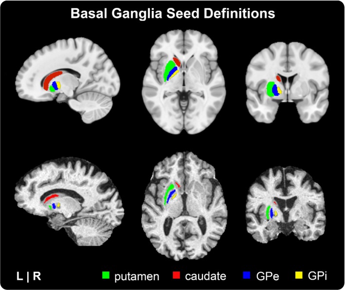
Resting-state data from 89 right-handed subjects were downloaded from the Parkinson's Progression Markers Initiative database. Subjects included 12 older healthy controls ("OHC"), 42 PD patients without speech impairment ("PDN"), and 35 PD subjects with speech impairment ("PDSI"). Subjects were assigned to PDN and PDSI groups based on the Movement Disorders Society Unified Parkinson's Disease Rating Scale (MDS-UPDRS) Part III speech item scores ("0" vs. "1-4"). Whole-brain functional connectivity was calculated for four basal ganglia seeds in each hemisphere: putamen, caudate, external globus pallidus (GPe), and internal globus pallidus (GPi). For each seed region, group-averaged connectivity maps were compared among OHC, PDN, and PDSI groups using a multivariate ANCOVA controlling for the effects of age and sex. Subsequent planned pairwise t-tests were performed to determine differences between the three groups using a voxel-wise threshold of p < 0.001 and cluster-extent threshold of 272 mm (FWE<0.05).
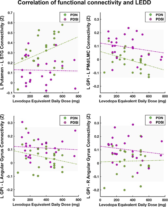
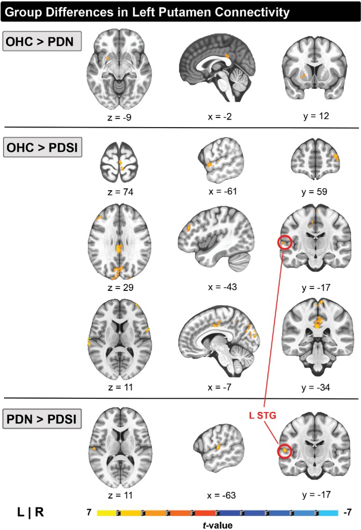
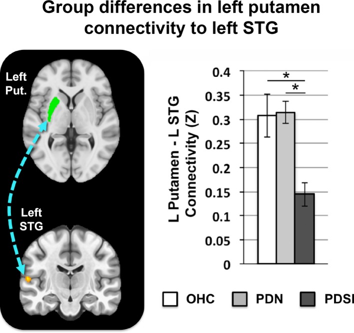
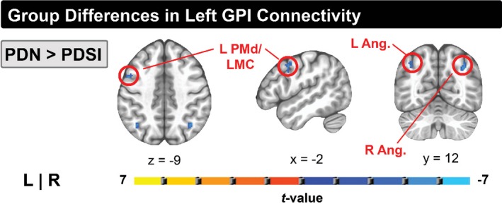
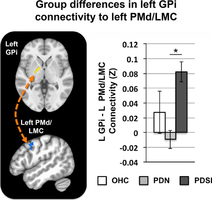
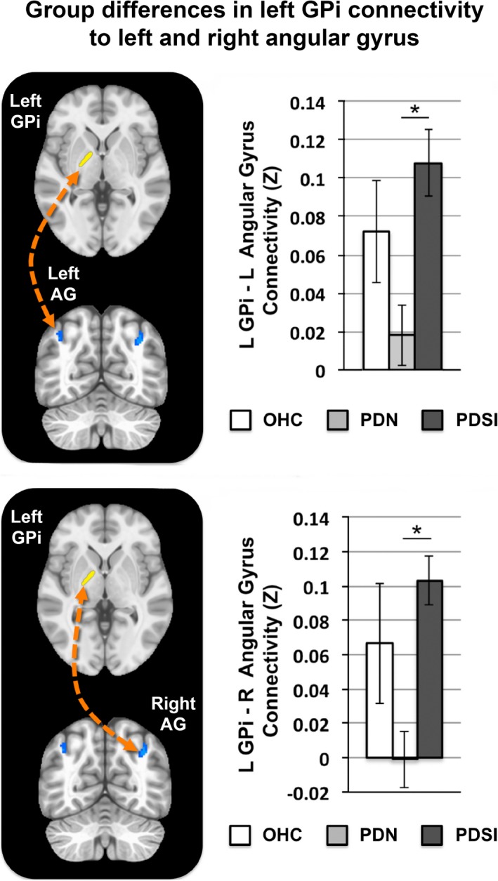
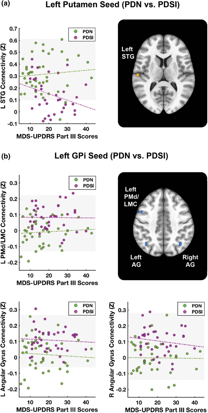
In comparison with OHCs, both PDN and PDSI groups demonstrated significant differences in cortical connectivity with bilateral putamen, bilateral GPe, and right caudate. Compared to the PDN group, the PDSI subjects demonstrated significant differences in cortical connectivity with left putamen and left GPi. PDSI subjects had lower connectivity between the left putamen and left superior temporal gyrus compared to PDN. In addition, PDSI subjects had greater connectivity between left GPi and three cortical regions: left dorsal premotor/laryngeal motor cortex, left angular gyrus, and right angular gyrus.
The present findings suggest that speech impairment in PD is associated with altered cortical connectivity with left putamen and left GPi.
帕金森病(PD)患者的言语障碍普遍存在,对生活有重大影响。然而,对于基底神经节和皮质之间的功能连接与 PD 言语障碍(PDSI)之间的关系知之甚少。基底神经节核团的全脑静息状态连接分析可以扩展对 PDSI 病理生理学的理解。
从帕金森进展标志物倡议数据库中下载了 89 名右利手受试者的静息态数据。受试者包括 12 名老年健康对照者(“OHC”)、42 名无言语障碍的 PD 患者(“PDN”)和 35 名有言语障碍的 PD 受试者(“PDSI”)。根据运动障碍协会统一帕金森病评定量表(MDS-UPDRS)第三部分言语项目评分(“0”与“1-4”),将受试者分为 PDN 和 PDSI 组。在每个半球的四个基底神经节种子中计算全脑功能连接:壳核、尾状核、外苍白球(GPe)和内苍白球(GPi)。对于每个种子区域,使用多元方差分析(ANCOVA)在控制年龄和性别影响的情况下比较 OHC、PDN 和 PDSI 组的组平均连接图。随后使用体素-wise 阈值 p < 0.001 和聚类-extent 阈值 272mm(FWE<0.05)进行了计划的两两 t 检验,以确定三组之间的差异。
与 OHC 相比,PDN 和 PDSI 组双侧壳核、双侧 GPe 和右侧尾状核的皮质连接均有显著差异。与 PDN 组相比,PDSI 受试者左壳核和左苍白球的皮质连接有显著差异。与 PDN 相比,PDSI 患者左壳核与左颞上回之间的连接较低。此外,PDSI 患者左苍白球与三个皮质区域的连接较强:左背侧运动前区/喉运动皮质、左角回和右角回。
本研究结果表明,PD 患者的言语障碍与左壳核和左苍白球的皮质连接改变有关。