Laboratory of Epigenetics and Diseases, Department of Pharmacology and Toxicology, National Institute of Pharmaceutical Education and Research (NIPER) S.A.S, Nagar, India.
Sci Rep. 2018 Aug 16;8(1):12295. doi: 10.1038/s41598-018-30541-3.
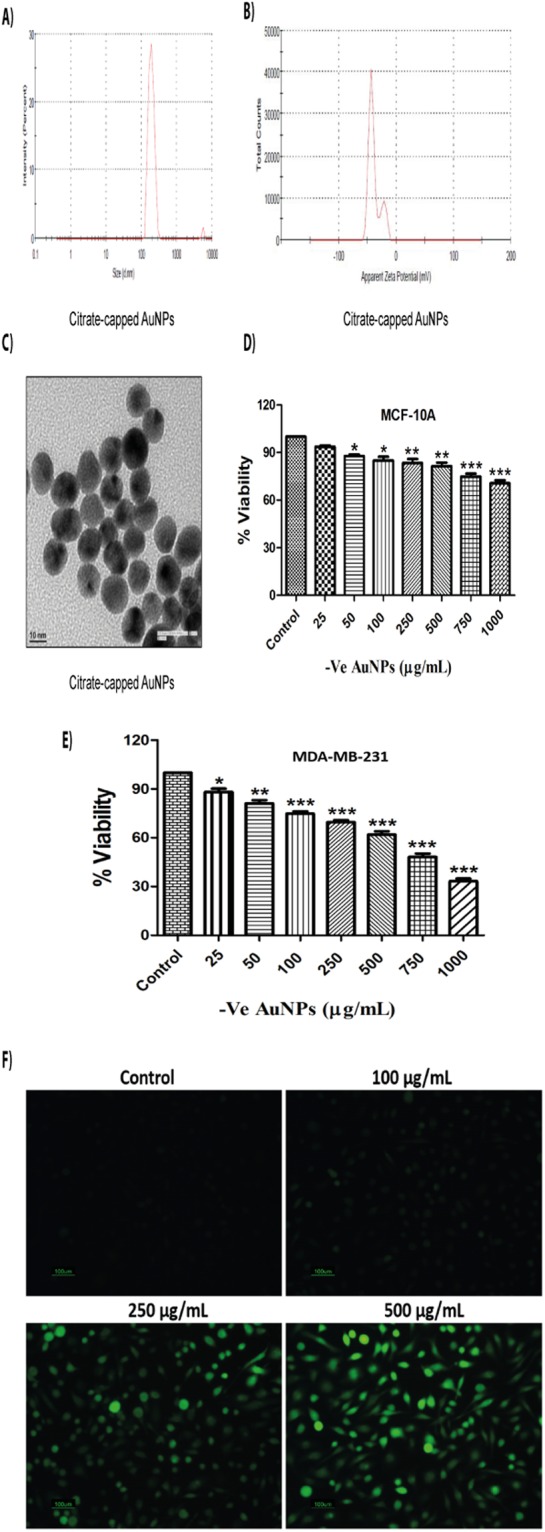
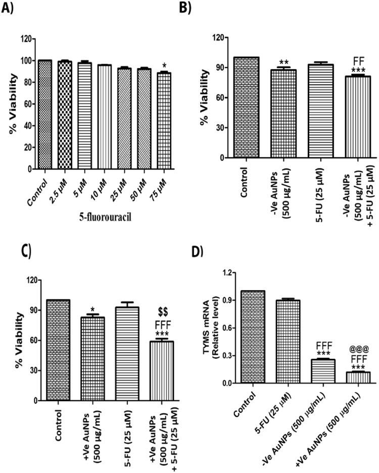
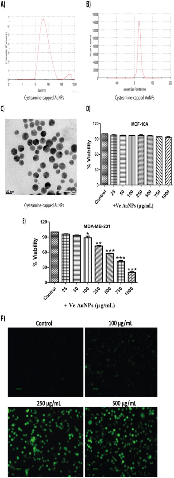
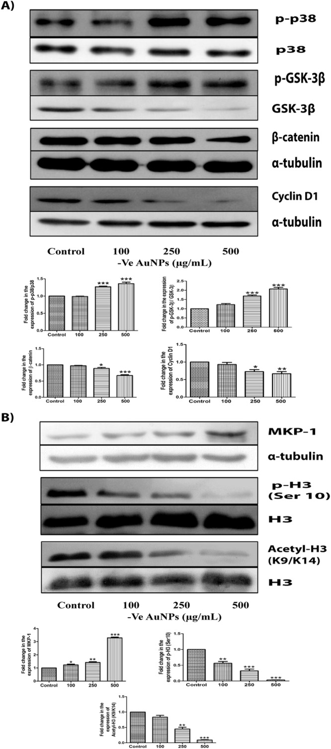
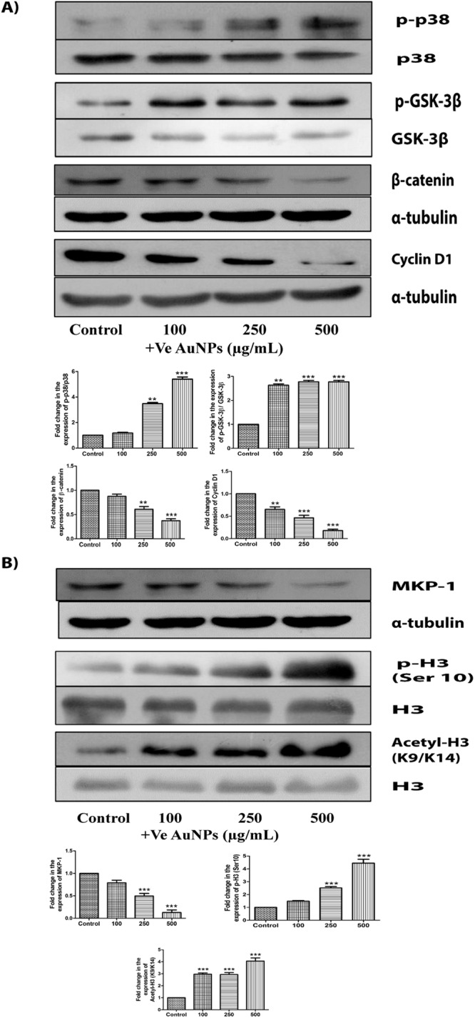
Gold nanoparticles (AuNPs) are used enormously in different cancers but very little is known regarding their molecular mechanism and surface charge role in the process of cell death. Here, we elucidate the molecular mechanism by which differentially charged AuNPs induce cytotoxicity in triple negative breast cancer (TNBC) cells. Cytotoxicity assay revealed that both negatively charged (citrate-capped) and positively charged (cysteamine-capped) AuNPs induced cell-death in a dose-dependent manner. We provide first evidence that AuNPs-induced oxidative stress alters Wnt signalling pathway in MDA-MB-231 and MDA-MB-468 cells. Although both differentially charged AuNPs induced cell death, the rate and mechanism involved in the process of cell death were different. Negatively charged AuNPs increased the expression of MKP-1, dephosphorylated and deacetylated histone H3 at Ser10 and K9/K14 residues respectively whereas, positively charged AuNPs decreased the expression of MKP-1, phosphorylated and acetylated histone H3 at Ser 10 and K9/K14 residues respectively. High-resolution transmission electron microscopy (HRTEM) studies revealed that AuNPs were localised in cytoplasm and mitochondria of MDA-MB-231 cells. Interestingly, AuNPs treatment makes MDA-MB-231 cells sensitive to 5-fluorouracil (5-FU) by decreasing the expression of thymidylate synthetase enzyme. This study highlights the role of surface charge (independent of size) in the mechanisms of toxicity and cell death.
金纳米粒子 (AuNPs) 在不同的癌症中被广泛应用,但对于它们在细胞死亡过程中的分子机制和表面电荷作用知之甚少。在这里,我们阐明了带不同电荷的 AuNPs 诱导三阴性乳腺癌 (TNBC) 细胞细胞毒性的分子机制。细胞毒性试验表明,带负电荷(柠檬酸封端)和带正电荷(半胱氨酸封端)的 AuNPs 均以剂量依赖的方式诱导细胞死亡。我们首次提供证据表明,AuNPs 诱导的氧化应激改变了 MDA-MB-231 和 MDA-MB-468 细胞中的 Wnt 信号通路。尽管带不同电荷的 AuNPs 均诱导细胞死亡,但细胞死亡过程中涉及的速率和机制不同。带负电荷的 AuNPs 增加了 MKP-1 的表达,使组蛋白 H3 在 Ser10 和 K9/K14 残基上去磷酸化和去乙酰化,而带正电荷的 AuNPs 则降低了 MKP-1 的表达,使组蛋白 H3 在 Ser10 和 K9/K14 残基上磷酸化和乙酰化。高分辨率透射电子显微镜 (HRTEM) 研究表明,AuNPs 定位于 MDA-MB-231 细胞的细胞质和线粒体中。有趣的是,AuNPs 处理通过降低胸苷酸合成酶的表达使 MDA-MB-231 细胞对 5-氟尿嘧啶 (5-FU) 敏感。这项研究强调了表面电荷(与尺寸无关)在毒性和细胞死亡机制中的作用。