Department of Biomedical Engineering, School of Medicine, Tsinghua University, Beijing 100084, China.
Department of Neurosurgery, Beijing Tiantan Hospital, Capital Medical University, Beijing, 100050 China.
Theranostics. 2018 Jul 16;8(15):4072-4085. doi: 10.7150/thno.25357. eCollection 2018.
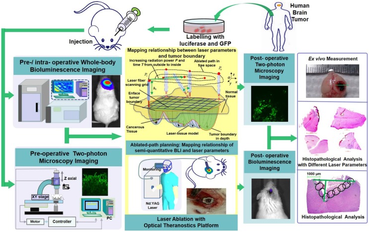
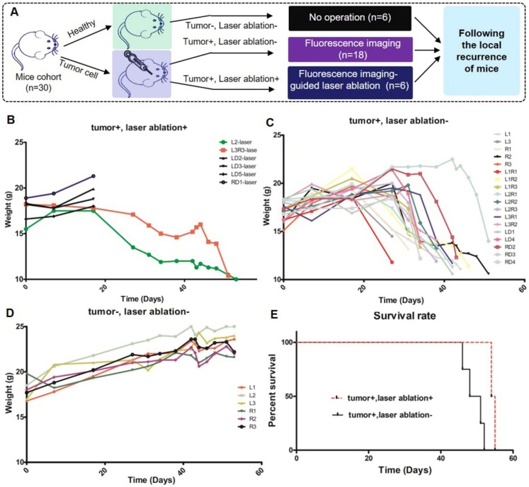
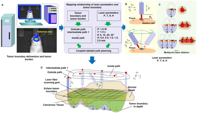
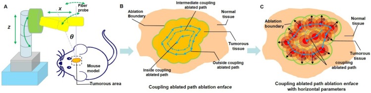
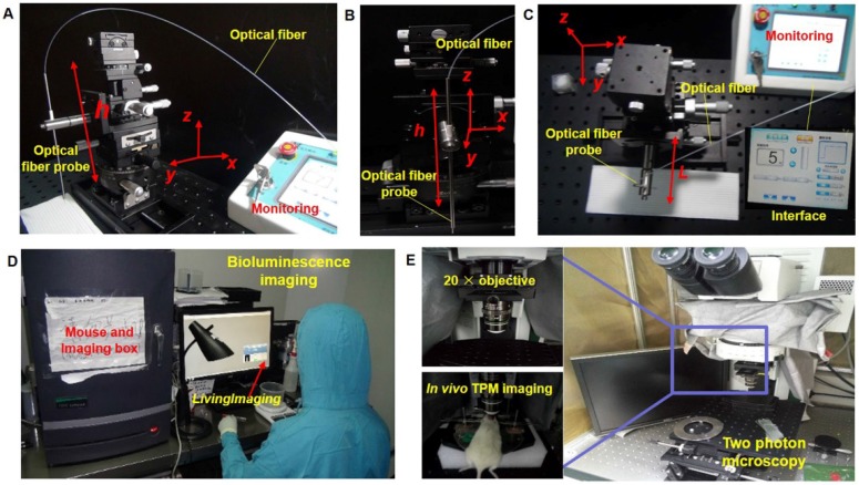
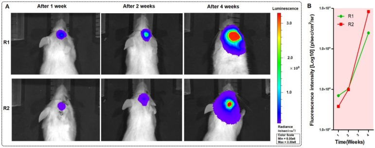
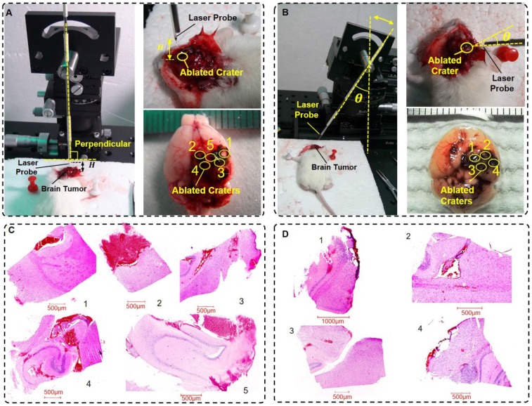
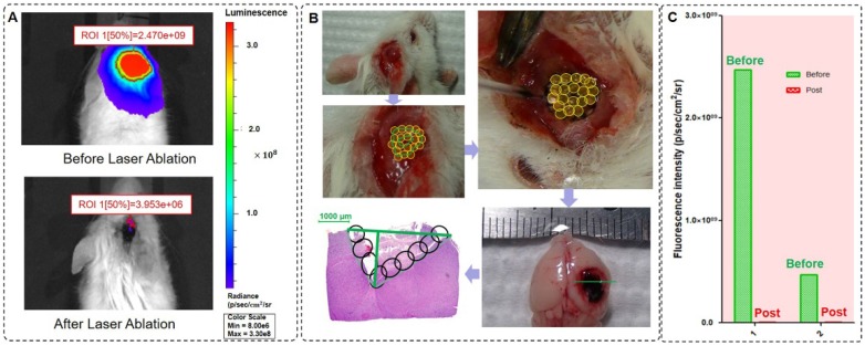
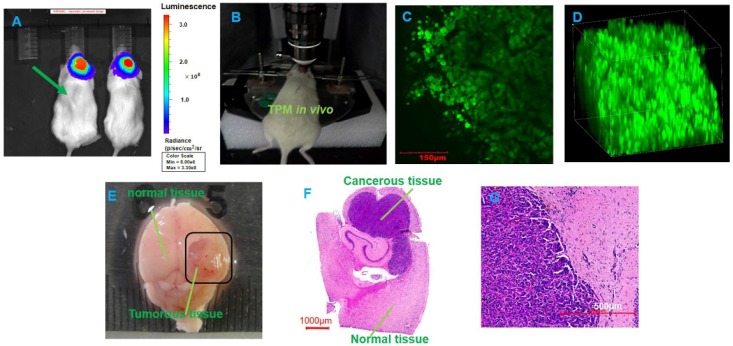
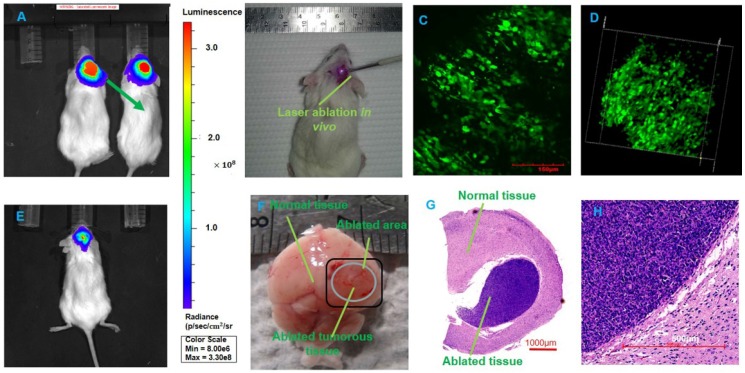
Brain tumor delineation and treatment are the main concerns of neurosurgeons in neurosurgical operations. Bridging the gap between imaging/diagnosis and treatment will provide great convenience for neurosurgeons. Here, we developed an optical theranostics platform that helps to delineate the boundary and quantitatively analyze glioblastoma multiforms (GBMs) with bioluminescence imaging (BLI) to guide laser ablation, and we imaged the GBM cells with two-photon microscopy (TPM) to visualize the laser ablation zone . Laser ablation, using the method of coupled ablated path planning with the guidance of BLI, was implemented for mouse brain tumors. The mapping relationship between semi-quantitative BLI and the laser ablation path was built through the quantitative tumor burden. The mapping was reflected through coupled ablated path planning. The BLI quantitatively and qualitatively evaluated treatment using laser ablation with the appropriate laser parameters and laser-tissue parameters. These parameters were measured after treatment. Furthermore, histopathological analysis of the brain tissue was conducted to compare the TPM images before and after laser ablation and to evaluate the results of laser ablation. The local recurrences were measured with three separate cohorts. The weights of all of the mice were measured during the experiment. Our BLI data show that the tumor cell numbers were significantly attenuated after treatment with the optical theranostics platform, and the delineation of GBM margins had clear views to guide the laser resection; the fluorescence intensity of GBMs quantitatively analyzed the rapid progression of GBMs. The laser-tissue parameters under guidance of multimodality imaging ranged between 1.0 mm and 0.1 mm. The accuracy of the laser ablation reached a submillimeter level, and the resection ratio reached more than 99% under the guidance of BLI. The histopathological sections were compared to TPM images, and the results demonstrated that these images highly coincided. The weight index and local recurrence results demonstrated that the therapeutic effect of the optical theranostics platform was significant. We propose an optical multimodality imaging-guided laser ablation theranostics platform for the treatment of GBMs in an intravital mouse model. The experimental results demonstrated that the integration of multimodality imaging can precisely guide laser ablation for the treatment of GBMs. This preclinical research provides a possibility for the precision treatment of GBMs. The study also provides some theoretical support for clinical research.
脑肿瘤的描绘和治疗是神经外科医生在神经外科手术中的主要关注点。弥合成像/诊断和治疗之间的差距将为神经外科医生提供极大的便利。在这里,我们开发了一种光学治疗平台,通过生物发光成像(BLI)帮助描绘胶质母细胞瘤多形性(GBM)的边界并进行定量分析,以指导激光消融,并通过双光子显微镜(TPM)对 GBM 细胞进行成像,以可视化激光消融区。使用 BLI 指导的耦合消融路径规划方法对小鼠脑肿瘤进行了激光消融。通过定量肿瘤负担建立了半定量 BLI 与激光消融路径之间的映射关系。通过耦合消融路径规划反映了映射关系。使用适当的激光参数和激光-组织参数对激光消融进行定量和定性评估。这些参数在治疗后进行测量。此外,还对脑组织进行了组织病理学分析,比较了激光消融前后的 TPM 图像,并评估了激光消融的结果。通过三个独立的队列测量局部复发情况。在实验过程中测量了所有小鼠的体重。我们的 BLI 数据表明,使用光学治疗平台治疗后,肿瘤细胞数量明显减少,GBM 边界的描绘清晰,可指导激光切除;GBM 的荧光强度定量分析了 GBM 的快速进展。在多模态成像指导下的激光-组织参数范围在 1.0 毫米到 0.1 毫米之间。在 BLI 指导下,激光消融的精度达到亚毫米级,切除率达到 99%以上。组织病理学切片与 TPM 图像进行了比较,结果表明这些图像高度吻合。体重指数和局部复发结果表明,光学治疗平台的治疗效果显著。我们提出了一种用于活体小鼠模型中 GBM 治疗的光学多模态成像引导激光消融治疗平台。实验结果表明,多模态成像的融合可以精确指导 GBM 的激光消融治疗。这项临床前研究为 GBM 的精准治疗提供了一种可能性。该研究也为临床研究提供了一些理论支持。