Department of Molecular Biology and Biotechnology, University of Kalyani,West Bengal, India.
Department of Botany, University of Kalyani, West Bengal, India.
PLoS One. 2018 Aug 22;13(8):e0202324. doi: 10.1371/journal.pone.0202324. eCollection 2018.
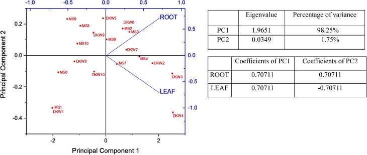
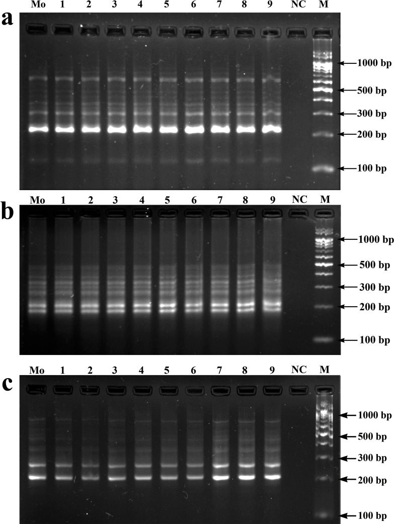
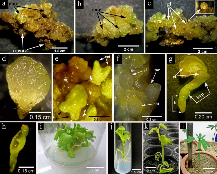
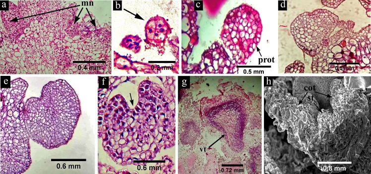
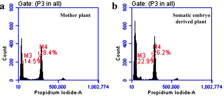
Induction of somatic embryogenesis and complete plantlet regeneration from callus culture of Hibiscus sabdariffa L. var. HS4288 has been made. Leaf and root explants were cultured on Murashige and Skoog (MS) and Driver-Kuniyuki Walnut (DKW) basal media supplemented with different concentrations of synthetic auxins and cytokinins. Root explants on DKW medium supplemented with 2.26μM 2, 4-Dichlorophenoxyacetic acid (2, 4-D) and 4.65μM kinetin (KIN) induced highest percentage (70%) of embryogenic calli. Average number of globular embryos per root derived callus produced within 6 weeks of culture initiation on MS media with different plant growth regulators (PGRs) ranged from 2.27±0.12 to 8.80±0.17 and that of cotyledonary embryos ranged from 0.00 to 2.53±0.20. On DKW medium comparatively more globular embryos (2.70±0.15 to 14.53±0.23) and cotyledonary embryos (0.00 to 8.90±0.17) were produced than that of MS medium. Regeneration of complete plantlets was highest (76.67%) when embryogenic calli with mature somatic embryos were grown on DKW medium containing 2.32μM KIN and 2.22μM 6-Benzyladenine (BA). Plants were primarily hardened in humidity, temperature and light controlled chamber and finally in a greenhouse showed 70% survival ability. Different stages of somatic embryogenesis process in the root derived embryogenic calli were elaborated in detail by morphological, histological and SEM study. The data were statistically analyzed by Duncan Multiple range test (p ≤ 0.05) and Principal component analysis (PCA). Flow cytometry and Inter-simple sequence repeats (ISSR) marker analysis confirmed that there was no genetic variation within the regenerated plants.
已成功诱导木槿(Hibiscus sabdariffa L. var. HS4288)愈伤组织的体细胞胚胎发生和完整植株再生。叶片和根外植体在 Murashige 和 Skoog (MS) 和 Driver-Kuniyuki 胡桃(DKW)基本培养基上培养,培养基中添加不同浓度的合成生长素和细胞分裂素。在添加 2.26μM 2,4-二氯苯氧乙酸(2,4-D)和 4.65μM 激动素(KIN)的 DKW 培养基上的根外植体诱导出最高比例(70%)的胚性愈伤组织。在不同植物生长调节剂(PGR)的 MS 培养基上,每块根衍生愈伤组织中产生的球状胚胎的平均数量在培养起始后 6 周内从 2.27±0.12 到 8.80±0.17,子叶胚的数量从 0.00 到 2.53±0.20。在 DKW 培养基上,与 MS 培养基相比,产生的球状胚胎(2.70±0.15 到 14.53±0.23)和子叶胚(0.00 到 8.90±0.17)更多。当含有 2.32μM KIN 和 2.22μM 6-苄基腺嘌呤(BA)的 DKW 培养基上生长成熟的体细胞胚的胚性愈伤组织时,完整植株的再生率最高(76.67%)。植物首先在湿度、温度和光照控制的室内进行硬化,最后在温室中显示出 70%的生存能力。通过形态学、组织学和扫描电镜研究详细阐述了根衍生胚性愈伤组织中不同阶段的体细胞胚胎发生过程。数据通过邓肯多重范围检验(p≤0.05)和主成分分析(PCA)进行了统计分析。流式细胞术和简单序列重复(ISSR)标记分析证实再生植物内没有遗传变异。