• 文献检索
  • 文档翻译
  • 深度研究
  • 学术资讯
  • Suppr Zotero 插件Zotero 插件
  • 邀请有礼
  • 套餐&价格
  • 历史记录
应用&插件
Suppr Zotero 插件Zotero 插件浏览器插件Mac 客户端Windows 客户端微信小程序
定价
高级版会员购买积分包购买API积分包
服务
文献检索文档翻译深度研究API 文档MCP 服务
关于我们
关于 Suppr公司介绍联系我们用户协议隐私条款
关注我们

Suppr 超能文献

核心技术专利:CN118964589B侵权必究
粤ICP备2023148730 号-1Suppr @ 2026

文献检索

告别复杂PubMed语法,用中文像聊天一样搜索,搜遍4000万医学文献。AI智能推荐,让科研检索更轻松。

立即免费搜索

文件翻译

保留排版,准确专业,支持PDF/Word/PPT等文件格式,支持 12+语言互译。

免费翻译文档

深度研究

AI帮你快速写综述,25分钟生成高质量综述,智能提取关键信息,辅助科研写作。

立即免费体验

超声微血管成像与传统超声鉴别BI-RADS 4类乳腺病变的对比研究

A comparative study on superb microvascular imaging and conventional ultrasonography in differentiating BI-RADS 4 breast lesions.

作者信息

Zhu Yi-Cheng, Zu Dao-Ming, Zhang Yuan, Shan Jun, Shi Xiu-Rong, Deng Shu-Hao, Jiang Quan

机构信息

Department of Ultrasound, Pudong New Area People's Hospital Affiliated to Shanghai University of Medicine and Health Sciences, Shanghai 201299, P.R. China.

Department of Minor Surgery, Pudong New Area People's Hospital Affiliated to Shanghai University of Medicine and Health Sciences, Shanghai 201299, P.R. China.

出版信息

Oncol Lett. 2019 Sep;18(3):3202-3210. doi: 10.3892/ol.2019.10603. Epub 2019 Jul 11.

DOI:10.3892/ol.2019.10603
PMID:31452797
原文链接:https://pmc.ncbi.nlm.nih.gov/articles/PMC6676576/
Abstract

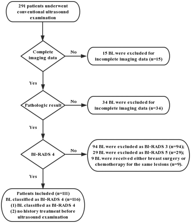
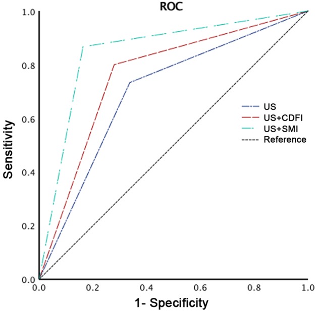
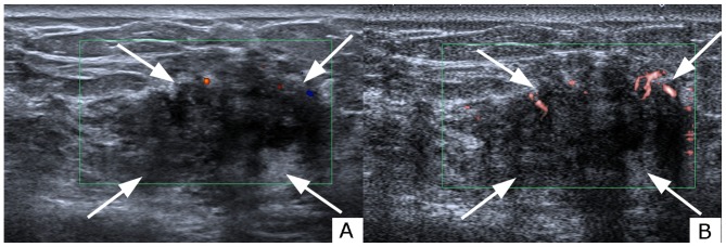
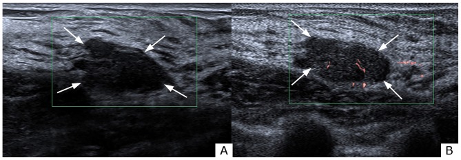
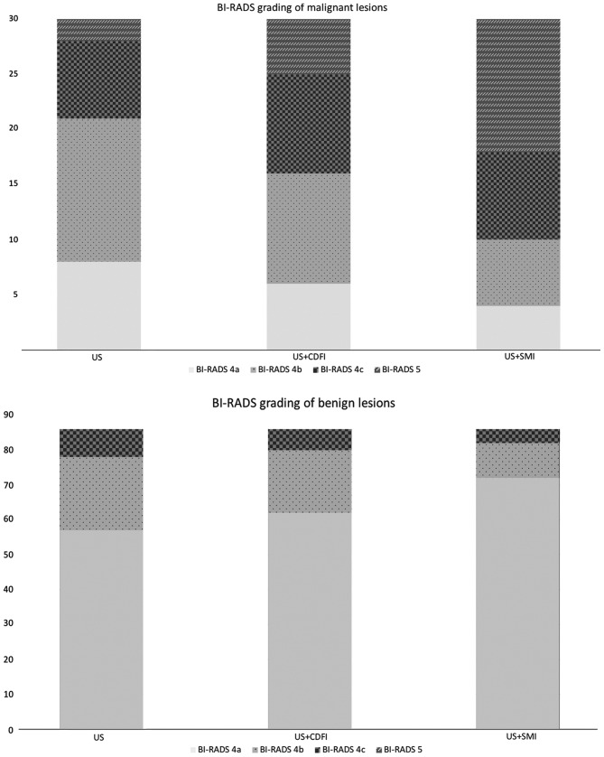
This prospective study aimed to explore the diagnostic value of superb microvascular imaging (SMI) in differentiating Breast Imaging Reporting and Data System (BI-RADS) 4 breast lesions compared with conventional ultrasonography (US). A total of 111 patients with 116 breast lesions underwent grayscale ultrasound (US), colour Doppler flow imaging (CDFI) and SMI breast imaging between February 2016 and May 2018. CDFI and SMI were performed to evaluate vascular quantity, morphology, and distribution characteristics. The detection of malignancy was compared between grayscale US alone, US + CDFI and US + SMI in terms of the BI-RADS stratification system. SMI was observed to be significantly more accurate in distinguishing malignant breast lesions (86.67%) compared with CDFI (80.00%) (P<0.001). Among malignant lesions, SMI detected 80.00% of those that contained ≥4 vessels, while CDFI only detected 56.67%. Penetrating and branching vessels were identified by SMI in 53.33% of malignant breast lesions and by CDFI in 10.00%. There was no significant difference in vascular distribution by SMI (P=0.094) and by CDFI (P=0.087). US + SMI was associated with higher sensitivity, specificity, and accuracy rates (86.67, 83.72 and 84.48%, respectively) compared with US + CDFI (80.00, 72.09 and 74.14%, respectively). The area under the curve values from receiver operating characteristic analysis of US + SMI, US + CDFI and US alone were 0.852 [95% confidence interval (CI): 0.768-0.936)] 0.760 (95% CI: 0.660-0.860), 0.698 (95% CI: 0.589-0.807), respectively (P<0.001). SMI yielded more detailed vascular information associated with malignant breast masses when compared with conventional US. Therefore, as an adjunct to grayscale, SMI exhibited a markedly improved diagnostic capability in distinguishing malignant and benign breast lesions, particularly those of BI-RADS category 4.

摘要

本前瞻性研究旨在探讨与传统超声(US)相比,超微血管成像(SMI)在鉴别乳腺影像报告和数据系统(BI-RADS)4类乳腺病变中的诊断价值。2016年2月至2018年5月期间,共有111例患有116个乳腺病变的患者接受了灰阶超声(US)、彩色多普勒血流成像(CDFI)和SMI乳腺成像检查。采用CDFI和SMI评估血管数量、形态及分布特征。依据BI-RADS分层系统,比较单纯灰阶US、US+CDFI以及US+SMI对恶性病变的检测情况。结果显示,与CDFI(80.00%)相比,SMI在鉴别乳腺恶性病变方面的准确性显著更高(86.67%)(P<0.001)。在恶性病变中,SMI检测出80.00%含有≥4支血管的病变,而CDFI仅检测出56.67%。SMI在53.33%的乳腺恶性病变中识别出穿透性和分支状血管,而CDFI仅识别出10.00%。SMI(P=0.094)和CDFI(P=0.087)在血管分布方面无显著差异。与US+CDFI(分别为80.00%、72.09%和74.14%)相比,US+SMI具有更高的敏感性、特异性和准确率(分别为86.67%、83.72%和84.48%)。US+SMI、US+CDFI及单纯US的受试者操作特征分析曲线下面积值分别为0.852[95%置信区间(CI):0.768-0.936]、0.760(95%CI:0.660-0.860)、0.698(95%CI:0.589-0.807)(P<0.001)。与传统US相比,SMI能提供与乳腺恶性肿块相关的更详细血管信息。因此,作为灰阶超声的辅助手段,SMI在鉴别乳腺良恶性病变,尤其是BI-RADS 4类病变时,诊断能力有显著提高。

https://cdn.ncbi.nlm.nih.gov/pmc/blobs/613f/6676576/c8baabd60b87/ol-18-03-3202-g04.jpg
https://cdn.ncbi.nlm.nih.gov/pmc/blobs/613f/6676576/bd7740e845b4/ol-18-03-3202-g00.jpg
https://cdn.ncbi.nlm.nih.gov/pmc/blobs/613f/6676576/096aabb589af/ol-18-03-3202-g01.jpg
https://cdn.ncbi.nlm.nih.gov/pmc/blobs/613f/6676576/1d589a549971/ol-18-03-3202-g02.jpg
https://cdn.ncbi.nlm.nih.gov/pmc/blobs/613f/6676576/fb29507eb146/ol-18-03-3202-g03.jpg
https://cdn.ncbi.nlm.nih.gov/pmc/blobs/613f/6676576/c8baabd60b87/ol-18-03-3202-g04.jpg

相似文献

1
A comparative study on superb microvascular imaging and conventional ultrasonography in differentiating BI-RADS 4 breast lesions.超声微血管成像与传统超声鉴别BI-RADS 4类乳腺病变的对比研究
Oncol Lett. 2019 Sep;18(3):3202-3210. doi: 10.3892/ol.2019.10603. Epub 2019 Jul 11.
2
A Prospective Study to Compare Superb Microvascular Imaging with Grayscale Ultrasound and Color Doppler Flow Imaging of Vascular Distribution and Morphology in Thyroid Nodules.一项对比甲状腺结节血管分布和形态的灰阶超声及彩色多普勒血流显像与超级微血管成像的前瞻性研究。
Med Sci Monit. 2018 Dec 19;24:9223-9231. doi: 10.12659/MSM.911695.
3
Diagnostic Performance of Superb Microvascular Imaging (SMI) Combined with Shear-Wave Elastography in Evaluating Breast Lesions.超声微血管成像(SMI)联合剪切波弹性成像对乳腺病变的诊断性能评估。
Med Sci Monit. 2018 Aug 26;24:5935-5942. doi: 10.12659/MSM.910399.
4
The Diagnostic Value of Superb Microvascular Imaging (SMI) in Detecting Blood Flow Signals of Breast Lesions: A Preliminary Study Comparing SMI to Color Doppler Flow Imaging.超微血管成像(SMI)在检测乳腺病变血流信号中的诊断价值:一项比较SMI与彩色多普勒血流成像的初步研究
Medicine (Baltimore). 2015 Sep;94(36):e1502. doi: 10.1097/MD.0000000000001502.
5
[Application value of superb microvascular imaging for diagnosis of different size renal solid tumors].[超微血管成像在不同大小肾实性肿瘤诊断中的应用价值]
Zhonghua Zhong Liu Za Zhi. 2021 Nov 23;43(11):1215-1221. doi: 10.3760/cma.j.cn112152-20191220-00832.
6
Superb microvascular imaging for evaluation of microvascularity in breast nodules compared with conventional Doppler imaging.与传统多普勒成像相比,用于评估乳腺结节微血管的超微血管成像技术卓越。
Quant Imaging Med Surg. 2023 Oct 1;13(10):7029-7040. doi: 10.21037/qims-23-136. Epub 2023 Sep 22.
7
A Prospective Study on the Value of Ultrasound Microflow Assessment to Distinguish Malignant from Benign Solid Breast Masses: Association between Ultrasound Parameters and Histologic Microvessel Densities.超声微血管流评估对鉴别良恶性实性乳腺肿块的价值的前瞻性研究:超声参数与组织微血管密度的相关性。
Korean J Radiol. 2019 May;20(5):759-772. doi: 10.3348/kjr.2018.0515.
8
Combined use of strain elastography and superb microvascular imaging with grayscale ultrasound according to the BI-RADS classification for differentiating benign from malignant solid breast masses.根据 BI-RADS 分类,联合应变弹性成像和灰阶超声 superb microvascular imaging 对良恶性实性乳腺肿块进行鉴别诊断。
Clin Hemorheol Microcirc. 2020;74(4):391-403. doi: 10.3233/CH-190693.
9
Superb Microvascular Imaging-A new vascular detecting ultrasonographic technique for avascular breast masses: A preliminary study.超微血管成像——一种用于无血管乳腺肿块的新型血管检测超声技术:一项初步研究。
Eur J Radiol. 2016 May;85(5):915-21. doi: 10.1016/j.ejrad.2015.12.011. Epub 2016 Jan 18.
10
Role of Superb Microvascular Imaging in Differentiating Between Malignant and Benign Solid Breast Masses.超级微血管成像在鉴别良恶性实性乳腺肿块中的作用。
Clin Breast Cancer. 2020 Dec;20(6):e786-e793. doi: 10.1016/j.clbc.2020.06.009. Epub 2020 Jul 3.

引用本文的文献

1
A preliminary study on the diagnostic value of contrast-enhanced ultrasound and micro-flow imaging for detecting blood flow signals in breast cancer patients.超声造影及微血流成像对乳腺癌患者血流信号检测的诊断价值初步研究
Gland Surg. 2024 Nov 30;13(11):2098-2106. doi: 10.21037/gs-24-264. Epub 2024 Nov 26.
2
Ultrasound-Based Noncontrast Microvascular Imaging for Evaluation of Breast Lesions: Imaging Techniques and Review of Diagnostic Criteria.基于超声的无造影剂微血管成像在乳腺病变评估中的应用:成像技术及诊断标准综述
Indian J Radiol Imaging. 2024 Mar 17;34(4):702-713. doi: 10.1055/s-0044-1782162. eCollection 2024 Oct.
3

本文引用的文献

1
A Prospective Study to Compare Superb Microvascular Imaging with Grayscale Ultrasound and Color Doppler Flow Imaging of Vascular Distribution and Morphology in Thyroid Nodules.一项对比甲状腺结节血管分布和形态的灰阶超声及彩色多普勒血流显像与超级微血管成像的前瞻性研究。
Med Sci Monit. 2018 Dec 19;24:9223-9231. doi: 10.12659/MSM.911695.
2
Diagnostic Performance of Superb Microvascular Imaging (SMI) Combined with Shear-Wave Elastography in Evaluating Breast Lesions.超声微血管成像(SMI)联合剪切波弹性成像对乳腺病变的诊断性能评估。
Med Sci Monit. 2018 Aug 26;24:5935-5942. doi: 10.12659/MSM.910399.
3
Comparison of Superb Microvascular Imaging With Color Flow and Power Doppler Imaging of Small Hepatocellular Carcinomas.
Consistency Analysis of CTLM Imaging and Mammography in the Diagnosis of Breast Tumor Lesions.
CTLM成像与乳腺钼靶摄影在乳腺肿瘤病变诊断中的一致性分析
J Healthc Eng. 2022 Mar 29;2022:5391636. doi: 10.1155/2022/5391636. eCollection 2022.
4
Integrating the incident angle of main vessel of breast nodule with BI-RADS can improve the efficacy of breast malignancy evaluation.将乳腺结节主血管的入射角与 BI-RADS 相结合可以提高乳腺恶性肿瘤评估的效果。
Sci Rep. 2024 Sep 6;14(1):20783. doi: 10.1038/s41598-024-71567-0.
5
Translating new breast ultrasound techniques into clinical practice: evaluating their intended uses and describing other unexpected uses for them.将新的乳腺超声技术应用于临床实践:评估其预期用途并描述其他意外用途。
Transl Breast Cancer Res. 2023 Jul 30;4:23. doi: 10.21037/tbcr-23-29. eCollection 2023.
6
Use of a commercial artificial intelligence-based mammography analysis software for improving breast ultrasound interpretations.利用商业人工智能乳腺分析软件提高乳腺超声解读水平。
Eur Radiol. 2024 Oct;34(10):6320-6331. doi: 10.1007/s00330-024-10718-3. Epub 2024 Apr 3.
7
Using a combination of superb microvascular imaging and other auxiliary ultrasound techniques to increase the accuracy of gray-scale ultrasound for breast masses.采用卓越的微血管成像技术和其他辅助超声技术相结合的方法,提高乳腺肿块灰阶超声的准确性。
BMC Cancer. 2024 Feb 16;24(1):224. doi: 10.1186/s12885-024-11981-9.
8
Multimodal ultrasound imaging for diagnostic differentiation of sclerosing adenosis from invasive ductal carcinoma.多模态超声成像用于硬化性腺病与浸润性导管癌的诊断鉴别
Quant Imaging Med Surg. 2024 Jan 3;14(1):877-887. doi: 10.21037/qims-23-524. Epub 2024 Jan 2.
9
A Novel Nomogram Based on Imaging Biomarkers of Shear Wave Elastography, Angio Planewave Ultrasensitive Imaging, and Conventional Ultrasound for Preoperative Prediction of Malignancy in Patients with Breast Lesions.基于剪切波弹性成像、血管平面波超敏成像和传统超声成像生物标志物的新型列线图用于术前预测乳腺病变患者的恶性肿瘤
Diagnostics (Basel). 2023 Feb 2;13(3):540. doi: 10.3390/diagnostics13030540.
10
Diagnostic Value of Superb Microvascular Imaging in Differentiating Benign and Malignant Breast Tumors: A Systematic Review and Meta-Analysis.超微血管成像在鉴别乳腺良恶性肿瘤中的诊断价值:一项系统评价与Meta分析
Diagnostics (Basel). 2022 Oct 31;12(11):2648. doi: 10.3390/diagnostics12112648.
微小肝细胞癌的微血管成像与彩色血流及能量多普勒成像的比较
J Ultrasound Med. 2018 Dec;37(12):2915-2924. doi: 10.1002/jum.14654. Epub 2018 Apr 23.
4
Angiogenesis - a crucial step in breast cancer growth, progression and dissemination by Raman imaging.血管生成——拉曼成像在乳腺癌生长、进展和扩散中的关键步骤。
Spectrochim Acta A Mol Biomol Spectrosc. 2018 Jun 5;198:338-345. doi: 10.1016/j.saa.2018.02.058. Epub 2018 Mar 2.
5
Superb microvascular imaging (SMI) compared with conventional ultrasound for evaluating thyroid nodules.与传统超声相比,超微血管成像(SMI)在评估甲状腺结节方面的应用
BMC Med Imaging. 2017 Dec 28;17(1):65. doi: 10.1186/s12880-017-0241-5.
6
Comparison of superb micro-vascular imaging (SMI) and conventional Doppler imaging techniques for evaluating testicular blood flow.用于评估睾丸血流的超微血管成像(SMI)与传统多普勒成像技术的比较。
J Med Ultrason (2001). 2018 Jul;45(3):443-452. doi: 10.1007/s10396-017-0847-9. Epub 2017 Dec 16.
7
Application of superb microvascular imaging in focal liver lesions.超微血管成像在肝脏局灶性病变中的应用。
World J Gastroenterol. 2017 Nov 21;23(43):7765-7775. doi: 10.3748/wjg.v23.i43.7765.
8
The utility of ultrasound superb microvascular imaging for evaluation of breast tumour vascularity: comparison with colour and power Doppler imaging regarding diagnostic performance.超声超微血管成像在评估乳腺肿瘤血管方面的应用:与彩色多普勒成像和能量多普勒成像诊断性能的比较
Clin Radiol. 2018 Mar;73(3):304-311. doi: 10.1016/j.crad.2017.10.006. Epub 2017 Nov 6.
9
Screening for Breast Cancer.乳腺癌筛查
Radiol Clin North Am. 2017 Nov;55(6):1145-1162. doi: 10.1016/j.rcl.2017.06.004.
10
Utility of BI-RADS Assessment Category 4 Subdivisions for Screening Breast MRI.乳腺影像报告和数据系统(BI-RADS)4类细分在乳腺磁共振成像筛查中的应用
AJR Am J Roentgenol. 2017 Jun;208(6):1392-1399. doi: 10.2214/AJR.16.16730.