• 文献检索
  • 文档翻译
  • 深度研究
  • 学术资讯
  • Suppr Zotero 插件Zotero 插件
  • 邀请有礼
  • 套餐&价格
  • 历史记录
应用&插件
Suppr Zotero 插件Zotero 插件浏览器插件Mac 客户端Windows 客户端微信小程序
定价
高级版会员购买积分包购买API积分包
服务
文献检索文档翻译深度研究API 文档MCP 服务
关于我们
关于 Suppr公司介绍联系我们用户协议隐私条款
关注我们

Suppr 超能文献

核心技术专利:CN118964589B侵权必究
粤ICP备2023148730 号-1Suppr @ 2026

文献检索

告别复杂PubMed语法,用中文像聊天一样搜索,搜遍4000万医学文献。AI智能推荐,让科研检索更轻松。

立即免费搜索

文件翻译

保留排版,准确专业,支持PDF/Word/PPT等文件格式,支持 12+语言互译。

免费翻译文档

深度研究

AI帮你快速写综述,25分钟生成高质量综述,智能提取关键信息,辅助科研写作。

立即免费体验

胸膜CT与标准X线摄影的对比解读

Comparative Interpretation of CT and Standard Radiography of the Pleura.

作者信息

Ilsen Bart, Vandenbroucke Frederik, Beigelman-Aubry Cathérine, Brussaard Carola, de Mey Johan

机构信息

UZ Brussel, BE.

出版信息

J Belg Soc Radiol. 2016 Nov 19;100(1):106. doi: 10.5334/jbr-btr.1229.

DOI:10.5334/jbr-btr.1229
PMID:30151497
原文链接:https://pmc.ncbi.nlm.nih.gov/articles/PMC6100645/
Abstract

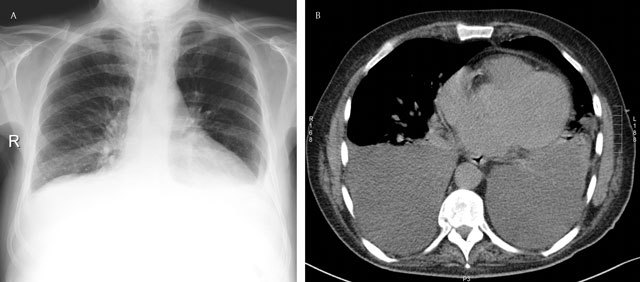
Many diseases affect the pleural space in both adults and children, including common diseases such as pneumonia, cancer and heart failure. Pleural effusion is the most common manifestation of pleural disease, and it is often a secondary effect of another disease process. Imaging plays a crucial role in the management of pleural disease. Chest radiography often remains the first examination in the assessment of these patients. Depending on the clinical context, the optimal imaging technique for further evaluation might be computed tomography (CT), ultrasound (US), or magnetic resonance (MR).

摘要

许多疾病会影响成人和儿童的胸膜腔,包括肺炎、癌症和心力衰竭等常见疾病。胸腔积液是胸膜疾病最常见的表现,它通常是另一种疾病过程的继发效应。影像学在胸膜疾病的管理中起着至关重要的作用。胸部X线摄影通常仍是评估这些患者的首要检查。根据临床情况,进一步评估的最佳影像学技术可能是计算机断层扫描(CT)、超声(US)或磁共振(MR)。

https://cdn.ncbi.nlm.nih.gov/pmc/blobs/032a/6100645/22db15b458bf/jbsr-100-1-1229-g14.jpg
https://cdn.ncbi.nlm.nih.gov/pmc/blobs/032a/6100645/486fff44c092/jbsr-100-1-1229-g1.jpg
https://cdn.ncbi.nlm.nih.gov/pmc/blobs/032a/6100645/2b384b235e64/jbsr-100-1-1229-g2.jpg
https://cdn.ncbi.nlm.nih.gov/pmc/blobs/032a/6100645/abf5c654a095/jbsr-100-1-1229-g3.jpg
https://cdn.ncbi.nlm.nih.gov/pmc/blobs/032a/6100645/8d01d85d5b84/jbsr-100-1-1229-g4.jpg
https://cdn.ncbi.nlm.nih.gov/pmc/blobs/032a/6100645/24256b538cd3/jbsr-100-1-1229-g5.jpg
https://cdn.ncbi.nlm.nih.gov/pmc/blobs/032a/6100645/3599cd3c1304/jbsr-100-1-1229-g6.jpg
https://cdn.ncbi.nlm.nih.gov/pmc/blobs/032a/6100645/fc49c2f39cfc/jbsr-100-1-1229-g7.jpg
https://cdn.ncbi.nlm.nih.gov/pmc/blobs/032a/6100645/6c5b044dd091/jbsr-100-1-1229-g8.jpg
https://cdn.ncbi.nlm.nih.gov/pmc/blobs/032a/6100645/479f852e8302/jbsr-100-1-1229-g9.jpg
https://cdn.ncbi.nlm.nih.gov/pmc/blobs/032a/6100645/035351f4f7e1/jbsr-100-1-1229-g10.jpg
https://cdn.ncbi.nlm.nih.gov/pmc/blobs/032a/6100645/555b5741e087/jbsr-100-1-1229-g11.jpg
https://cdn.ncbi.nlm.nih.gov/pmc/blobs/032a/6100645/9138d6f8ef08/jbsr-100-1-1229-g12.jpg
https://cdn.ncbi.nlm.nih.gov/pmc/blobs/032a/6100645/984b3814ccb4/jbsr-100-1-1229-g13.jpg
https://cdn.ncbi.nlm.nih.gov/pmc/blobs/032a/6100645/22db15b458bf/jbsr-100-1-1229-g14.jpg

相似文献

1
Comparative Interpretation of CT and Standard Radiography of the Pleura.胸膜CT与标准X线摄影的对比解读
J Belg Soc Radiol. 2016 Nov 19;100(1):106. doi: 10.5334/jbr-btr.1229.
2
MR imaging of the pleura and chest wall.胸膜和胸壁的磁共振成像。
Magn Reson Imaging Clin N Am. 2000 Feb;8(1):125-41.
3
[Malignant pleural diseases: diagnosis and treatment].[恶性胸膜疾病:诊断与治疗]
Medicina (Kaunas). 2008;44(12):929-35.
4
A Simple Method for Differentiating Complicated Parapneumonic Effusion/Empyema from Parapneumonic Effusion Using the Split Pleura Sign and the Amount of Pleural Effusion on Thoracic CT.一种利用胸腔CT上的胸膜分裂征和胸腔积液量来鉴别复杂性类肺炎性胸腔积液/脓胸与类肺炎性胸腔积液的简单方法。
PLoS One. 2015 Jun 15;10(6):e0130141. doi: 10.1371/journal.pone.0130141. eCollection 2015.
5
Radiology in pleural disease: state of the art.胸膜疾病的放射学:最新进展
Respirology. 2004 Aug;9(3):300-12. doi: 10.1111/j.1440-1843.2004.00599.x.
6
Radiographic and computed tomography (CT) imaging of complex pleural disease.复杂胸膜疾病的放射学及计算机断层扫描(CT)成像
Crit Rev Diagn Imaging. 1997;38(1):1-58.
7
Imaging the pleura: sonography, CT, and MR imaging.胸膜成像:超声检查、CT及磁共振成像
AJR Am J Roentgenol. 1991 Jun;156(6):1145-53. doi: 10.2214/ajr.156.6.2028857.
8
Asbestos: when the dust settles an imaging review of asbestos-related disease.石棉:尘埃落定之时——石棉相关疾病的影像学综述
Radiographics. 2002 Oct;22 Spec No:S167-84. doi: 10.1148/radiographics.22.suppl_1.g02oc10s167.
9
Imaging of pleural diseases.胸膜疾病的影像学检查
Monaldi Arch Chest Dis. 1996 Apr;51(2):138-44.
10
[Imaging of pleural diseases: evaluation of imaging methods based on chest radiography].[胸膜疾病的影像学检查:基于胸部X线摄影的成像方法评估]
Tuberk Toraks. 2017 Mar;65(1):41-55. doi: 10.5578/tt.47546.

引用本文的文献

1
Predictive Role of Venous Drainage and Lesion Margins in Bronchopulmonary Sequestration Classification.静脉引流和病变边缘在肺隔离症分类中的预测作用
J Clin Med. 2025 Apr 27;14(9):3018. doi: 10.3390/jcm14093018.
2
Effect of deep learning-based assistive technology use on chest radiograph interpretation by emergency department physicians: a prospective interventional simulation-based study.基于深度学习的辅助技术应用对急诊科医生胸部X光片解读的影响:一项基于前瞻性介入模拟的研究。
BMC Med Inform Decis Mak. 2021 Nov 8;21(1):311. doi: 10.1186/s12911-021-01679-4.

本文引用的文献

1
Clinical consequences of asbestos-related diffuse pleural thickening: A review.石棉相关弥漫性胸膜增厚的临床后果:综述。
J Occup Med Toxicol. 2008 Sep 8;3:20. doi: 10.1186/1745-6673-3-20.
2
Digital radiography with dual-energy subtraction: improved evaluation of cardiac calcification.采用双能减影的数字X线摄影:改善心脏钙化的评估
AJR Am J Roentgenol. 2004 Nov;183(5):1233-8. doi: 10.2214/ajr.183.5.1831233.
3
The shape of a pleural effusion.胸腔积液的形态。
Br Med J. 1963 Feb 16;1(5328):436-7. doi: 10.1136/bmj.1.5328.436.
4
Asbestos-related benign pleural disease.石棉相关的良性胸膜疾病
Clin Radiol. 2000 Jun;55(6):422-32. doi: 10.1053/crad.2000.0450.
5
Localized benign fibrous tumors of the pleura.
AJR Am J Roentgenol. 1997 Sep;169(3):683-6. doi: 10.2214/ajr.169.3.9275877.
6
Imaging of the pleura.胸膜的影像学检查。
Radiology. 1993 Feb;186(2):297-309. doi: 10.1148/radiology.186.2.8421723.
7
Pleural effusion: explanation of some typical appearances.胸腔积液:一些典型表现的解读
AJR Am J Roentgenol. 1982 Nov;139(5):899-904. doi: 10.2214/ajr.139.5.899.
8
Recognition of pleural effusion on supine radiographs: how much fluid is required?仰卧位X线片上胸腔积液的识别:需要多少液体?
AJR Am J Roentgenol. 1984 Jan;142(1):59-64. doi: 10.2214/ajr.142.1.59.
9
Differentiating lung abscess and empyema: radiography and computed tomography.肺脓肿与脓胸的鉴别:X线摄影与计算机断层扫描
AJR Am J Roentgenol. 1983 Jul;141(1):163-7. doi: 10.2214/ajr.141.1.163.
10
Asbestos-related pleural thickening: effect of threshold criteria on interpretation.石棉相关的胸膜增厚:阈值标准对解读的影响
Radiology. 1984 Sep;152(3):569-73. doi: 10.1148/radiology.152.3.6463238.