Institute of Process Engineering, Chair of Mechanical Process Engineering, Otto-von-Guericke University, Universitätsplatz 2, 39106 Magdeburg, Germany.
Institute of Medical Psychology, Otto-von-Guericke University, Leipziger Str. 44, 39120 Magdeburg, Germany.
Int J Mol Sci. 2018 Sep 3;19(9):2613. doi: 10.3390/ijms19092613.
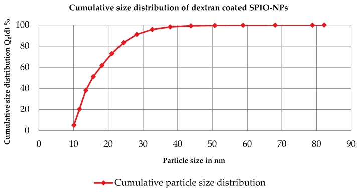
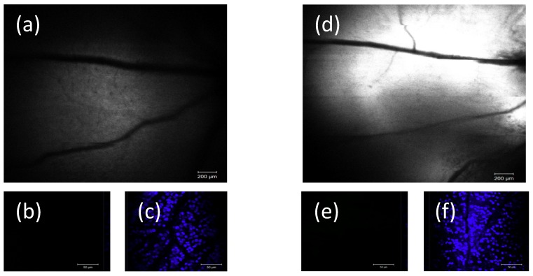
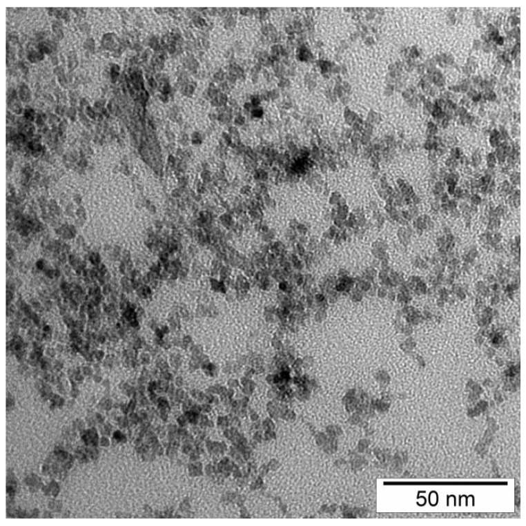
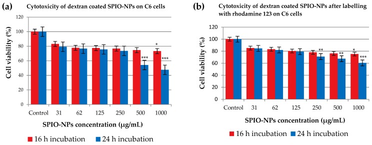
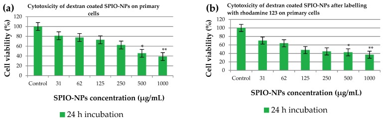
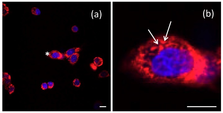
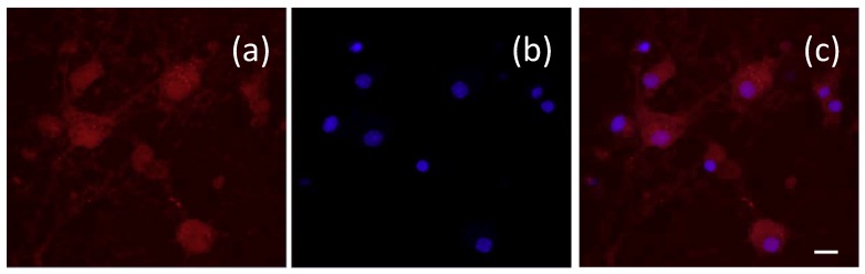
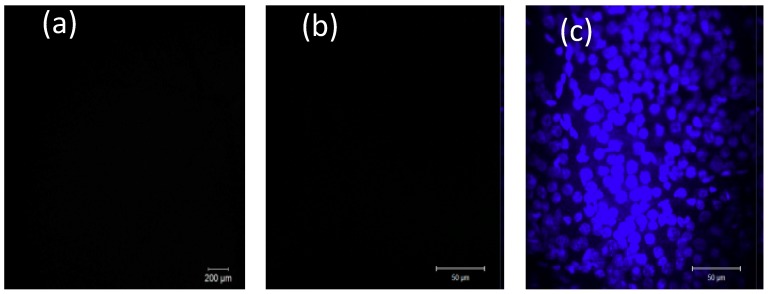
Superparamagnetic iron oxide nanoparticles (SPIO-NPs) have great potential to be used in different pharmaceutical applications, due to their unique and versatile physical and chemical properties. The aim of this study was to quantify in vitro cytotoxicity of dextran 70,000-coated SPIO-NPs labelled/unlabelled with rhodamine 123, in C6 glioma cells and primary hippocampal neural cells. In addition, we analyzed the in vitro and in vivo cellular uptake of labelled SPIO-NPs. The nanoparticles, with average size of 10⁻50 nm and polydispersity index of 0.37, were synthesized using Massart's co-precipitation method. The concentration-dependent cytotoxicity was quantified by using tetrazolium dye 3-(4,5-dimethylthiazol-2-yl)-2,5-diphenyltetrazolium bromide (MTT). Intracellular localization of SPIO-NPs was detected by confocal laser microscopy. In vivo confocal neuroimaging (ICON) was performed on male Wistar rats after intravitreal injection followed by ex vivo retina whole mount analysis. When used for in vitro testing concentrations in the range of diagnostic and therapeutic dosages, SPIO-NPs proved to be non-cytotoxic on C6 glioma cells for up to 24 h incubation time. The hippocampal cell culture also did not show impaired viability at low doses after 24 h incubation. Our results indicate that our dextran-coated SPIO-NPs have the potential for in vivo drug delivery applications.
超顺磁性氧化铁纳米颗粒(SPIO-NPs)由于其独特且多功能的物理和化学性质,在不同的药物应用中具有巨大的潜力。本研究旨在量化体外细胞毒性德赛醇 70,000 涂层 SPIO-NPs 标记/未标记 with rhodamine 123,在 C6 神经胶质瘤细胞和原代海马神经元细胞中。此外,我们还分析了标记 SPIO-NPs 的体外和体内细胞摄取。纳米颗粒的平均粒径为 10-50nm,多分散指数为 0.37,采用 Massart 的共沉淀法合成。使用四唑染料 3-(4,5-二甲基噻唑-2-基)-2,5-二苯基四唑溴盐(MTT)量化浓度依赖性细胞毒性。通过共聚焦激光显微镜检测 SPIO-NPs 的细胞内定位。在玻璃体腔注射后进行体内共聚焦神经成像(ICON),并对雄性 Wistar 大鼠进行视网膜全层分析。当用于体外测试浓度范围在诊断和治疗剂量范围内时,SPIO-NPs 在长达 24 小时的孵育时间内对 C6 神经胶质瘤细胞无细胞毒性。低剂量孵育 24 小时后,海马细胞培养也未显示出活力受损。我们的结果表明,我们的葡聚糖涂层 SPIO-NPs 具有体内药物输送应用的潜力。