Sarica Alessia, Vasta Roberta, Novellino Fabiana, Vaccaro Maria Grazia, Cerasa Antonio, Quattrone Aldo
Neuroscience Centre, Magna Graecia University, Catanzaro, Italy.
Neuroimaging Research Unit, Institute of Molecular Bioimaging and Physiology, National Research Council, Catanzaro, Italy.
Front Neurosci. 2018 Aug 21;12:576. doi: 10.3389/fnins.2018.00576. eCollection 2018.
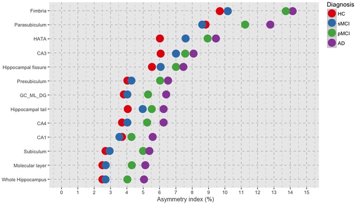
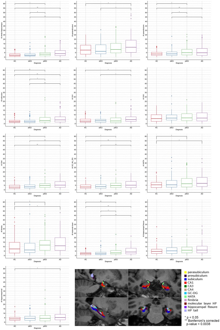
It is well-known that the hippocampus presents significant asymmetry in Alzheimer's disease (AD) and that difference in volumes between left and right exists and varies with disease progression. However, few works investigated whether the asymmetry degree of subfields of hippocampus changes through the continuum from Mild Cognitive Impairment (MCI) to AD. Thus, aim of the present work was to evaluate the Asymmetry Index (AI) of hippocampal substructures as possible MRI biomarkers of Dementia. Moreover, we aimed to assess whether the subfields presented peculiar differences between left and right hemispheres. We also investigated the relationship between the asymmetry magnitude in hippocampal subfields and the decline of verbal memory as assessed by Rey's auditory verbal learning test (RAVLT). Four-hundred subjects were selected from ADNI, equally divided into healthy controls (HC), AD, stable MCI (sMCI), and progressive MCI (pMCI). The structural baseline T1s were processed with FreeSurfer 6.0 and volumes of whole hippocampus (WH) and 12 subfields were extracted. The AI was calculated as: (|Left-Right|/(Left+Right))100. ANCOVA was used for evaluating AI differences between diagnoses, while paired -test was applied for assessing changes between left and right volumes, separately for each group. Partial correlation was performed for exploring relationship between RAVLT summary scores (Immediate, Learning, Forgetting, Percent Forgetting) and hippocampal substructures AI. The statistical threshold was Bonferroni corrected < 0.05/13 = 0.0038. We found a general trend of increased degree of asymmetry with increasing severity of diagnosis. Indeed, AD presented the higher magnitude of asymmetry compared with HC, sMCI and pMCI, in the WH (AI mean 5.13 ± 4.29 SD) and in each of its twelve subfields. Moreover, we found in AD a significant negative correlation ( = -0.33, = 0.00065) between the AI of parasubiculum (mean 12.70 ± 9.59 SD) and the RAVLT Learning score (mean 1.70 ± 1.62 SD). Our findings showed that hippocampal subfields AI varies differently among the four groups HC, sMCI, pMCI, and AD. Moreover, we found-for the first time-that hippocampal substructures had different sub-patterns of lateralization compared with the whole hippocampus. Importantly, the severity in learning rate was correlated with pathological high degree of asymmetry in parasubiculum of AD patients.
众所周知,海马体在阿尔茨海默病(AD)中存在显著不对称性,左右海马体体积存在差异且随疾病进展而变化。然而,很少有研究探讨海马体子区域的不对称程度在从轻度认知障碍(MCI)到AD的连续过程中是否发生变化。因此,本研究的目的是评估海马体亚结构的不对称指数(AI),将其作为痴呆症可能的MRI生物标志物。此外,我们旨在评估左右半球的子区域是否存在特殊差异。我们还研究了海马体子区域不对称程度与雷伊听觉词语学习测验(RAVLT)评估的言语记忆衰退之间的关系。从阿尔茨海默病神经成像计划(ADNI)中选取了400名受试者,平均分为健康对照组(HC)、AD组、稳定型MCI(sMCI)组和进展型MCI(pMCI)组。使用FreeSurfer 6.0对结构基线T1加权像进行处理,提取整个海马体(WH)和12个子区域的体积。AI的计算方法为:(|左 - 右|/(左 + 右))×100。采用协方差分析评估不同诊断组之间的AI差异,同时对每组分别应用配对t检验评估左右体积的变化。进行偏相关分析以探索RAVLT总结分数(即时、学习、遗忘、遗忘百分比)与海马体亚结构AI之间的关系。统计阈值经Bonferroni校正 < 0.05/13 = 0.0038。我们发现随着诊断严重程度的增加,不对称程度呈总体上升趋势。事实上,在WH(AI均值5.13 ± 4.29标准差)及其十二个子区域中,AD组的不对称程度高于HC组、sMCI组和pMCI组。此外,我们发现在AD组中,副下托的AI(均值12.70 ± 9.59标准差)与RAVLT学习分数(均值1.70 ± 1.62标准差)之间存在显著负相关(r = -0.33,p = 0.00065)。我们的研究结果表明,海马体子区域AI在HC、sMCI、pMCI和AD这四组中变化不同。此外,我们首次发现海马体亚结构与整个海马体相比具有不同的偏侧化子模式。重要的是,学习率的严重程度与AD患者副下托的病理性高度不对称相关。