Meng Hai-Jiang, Pi Yan-Ling, Liu Ke, Cao Na, Wang Yan-Qiu, Wu Yin, Zhang Jian
School of Kinesiology, Shanghai University of Sport, Shanghai, China.
School of Sports, Anqing Normal University, Anqing, China.
PeerJ. 2018 Aug 28;6:e5588. doi: 10.7717/peerj.5588. eCollection 2018.
Both motor imagery (MI) and motor execution (ME) can facilitate motor cortical excitability. Although cortical excitability is modulated by intracortical inhibitory and excitatory circuits in the human primary motor cortex, it is not clear which intracortical circuits determine the differences in corticospinal excitability between ME and MI.
We recruited 10 young healthy subjects aged 18-28 years (mean age: 22.1 ± 3.14 years; five women and five men) for this study. The experiment consisted of two sets of tasks involving grasp actions of the right hand: imagining and executing them. Corticospinal excitability and short-interval intracortical inhibition (SICI) were measured before the interventional protocol using transcranial magnetic stimulation (baseline), as well as at 0, 20, and 40 min (T0, T20, and T40) thereafter.
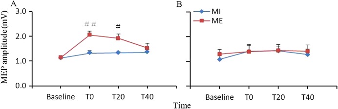
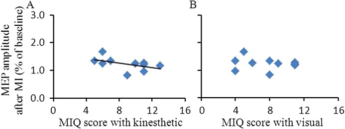
Facilitation of corticospinal excitability was significantly greater after ME than after MI in the right abductor pollicis brevis (APB) at T0 and T20 ( < 0.01 for T0, and < 0.05 for T20), but not in the first dorsal interosseous (FDI) muscle. On the other hand, no significant differences in SICI between ME and MI were found in the APB and FDI muscles. The facilitation of corticospinal excitability at T20 after MI correlated with the Movement Imagery Questionnaire (MIQ) scores for kinesthetic items ( = -0.646, = 0.044) but did not correlate with the MIQ scores for visual items ( = -0.265, = 0.458).
The present results revealed significant differences between ME and MI on intracortical excitatory circuits of the human motor cortex, suggesting that cortical excitability differences between ME and MI may be attributed to the activation differences of the excitatory circuits in the primary motor cortex.
运动想象(MI)和运动执行(ME)均能促进运动皮质兴奋性。尽管皮质兴奋性受人类初级运动皮质内皮质抑制性和兴奋性回路的调节,但尚不清楚哪些皮质内回路决定了ME和MI之间皮质脊髓兴奋性的差异。
我们招募了10名年龄在18 - 28岁的年轻健康受试者(平均年龄:22.1 ± 3.14岁;5名女性和5名男性)参与本研究。实验包括两组涉及右手抓握动作的任务:想象和执行这些动作。在干预方案前使用经颅磁刺激测量皮质脊髓兴奋性和短间隔皮质内抑制(SICI)(基线),以及此后的0、20和40分钟(T0、T20和T40)。
在T0和T20时,右侧拇短展肌(APB)在ME后皮质脊髓兴奋性的促进作用显著大于MI后(T0时P < 0.01,T20时P < 0.05),但在第一背侧骨间肌(FDI)中并非如此。另一方面,在APB和FDI肌肉中,ME和MI之间的SICI没有显著差异。MI后T20时皮质脊髓兴奋性的促进作用与运动想象问卷(MIQ)中动觉项目的得分相关(r = -0.646,P = 0.044),但与MIQ中视觉项目的得分不相关(r = -0.265,P = 0.458)。
本研究结果揭示了人类运动皮质内皮质兴奋性回路在ME和MI之间存在显著差异,表明ME和MI之间的皮质兴奋性差异可能归因于初级运动皮质兴奋性回路的激活差异。