C. and O. Vogt Institute for Brain Research, Medical Faculty, University Hospital Düsseldorf, Heinrich Heine University Düsseldorf, Düsseldorf, Germany.
Department of Neurology, The Affiliated Hospital of Southwest Medical University, Luzhou, China.
Brain Struct Funct. 2018 Dec;223(9):4169-4186. doi: 10.1007/s00429-018-1738-6. Epub 2018 Sep 5.
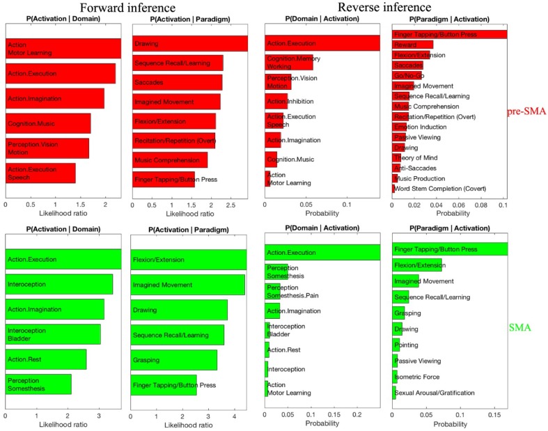
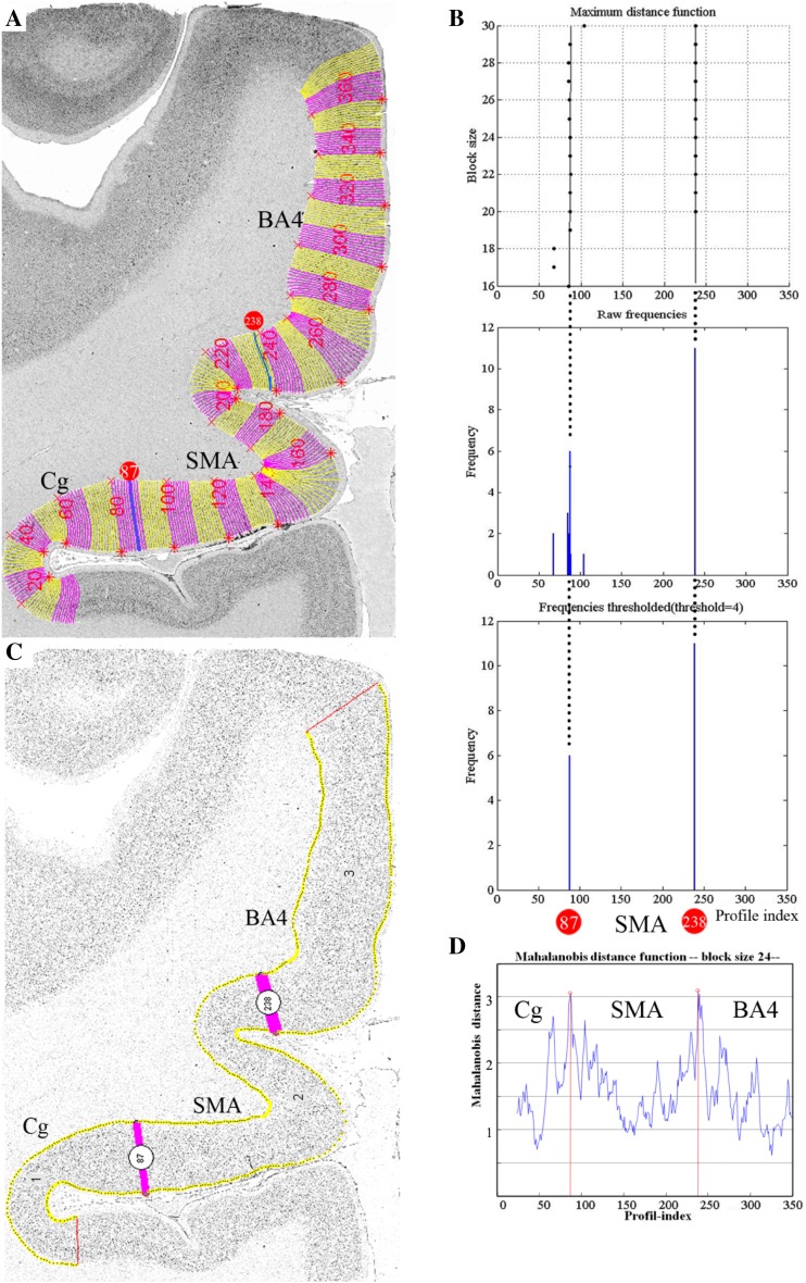
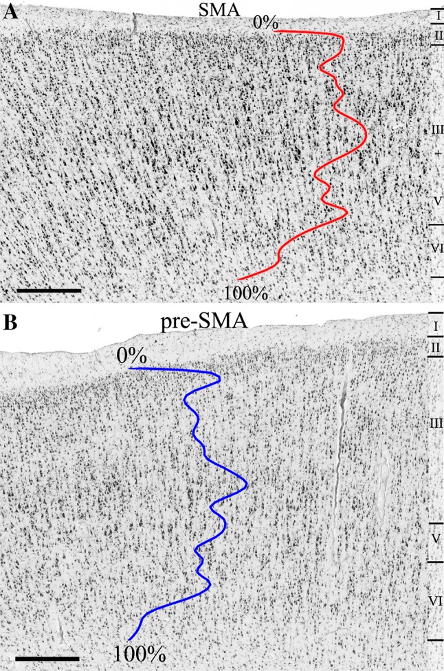
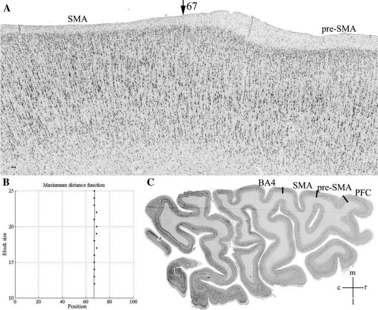
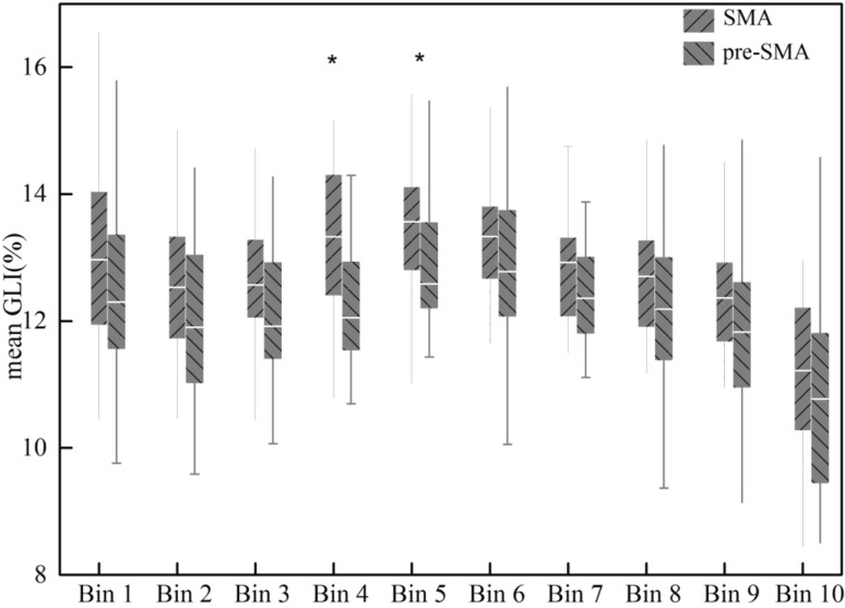
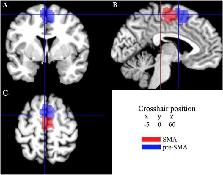
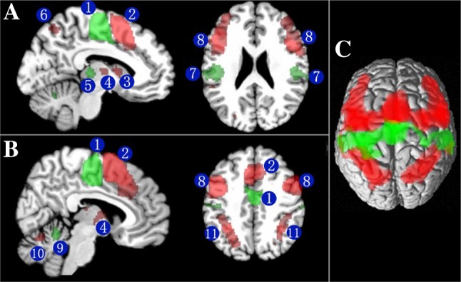
The dorsal mesial frontal cortex contains the supplementary motor area (SMA) and the pre-supplementary motor area (pre-SMA), which play an important role in action and cognition. Evidence from cytoarchitectonic, stimulation, and functional studies suggests structural and functional divergence between the two subregions. However, a microstructural map of these areas obtained in a representative sample of brains in a stereotaxic reference space is still lacking. In the present study we show that the dorsal mesial frontal motor cortex comprises two microstructurally different brain regions: area SMA and area pre-SMA. Area-specific cytoarchitectonic patterns were studied in serial histological sections stained for cell bodies of ten human postmortem brains. Borders of the two cortical areas were identified using image analysis and statistical features. The 3D reconstructed areas were transferred to a common reference space, and probabilistic maps were calculated by superimposing the individual maps. A coordinate-based meta-analysis of functional imaging data was subsequently performed using the two probabilistic maps as microstructurally defined seed regions. It revealed that areas SMA and pre-SMA were strongly co-activated with areas in precentral, supramarginal and superior frontal gyri, Rolandic operculum, thalamus, putamen and cerebellum. Both areas were related to motor functions, but area pre-SMA was involved in more complex processes such as learning, cognitive processes and perception. The here described subsequent analyses led to converging evidence supporting the microstructural, and functional segregation of areas SMA and pre-SMA, and maps will be made available to the scientific community to further elucidate the microstructural substrates of motor and cognitive control.
背内侧额上回包含补充运动区(SMA)和预备补充运动区(pre-SMA),它们在运动和认知中起着重要作用。来自细胞构筑、刺激和功能研究的证据表明这两个亚区在结构和功能上存在差异。然而,在一个代表性的立体参考空间的大脑样本中,仍然缺乏这些区域的微观结构图谱。在本研究中,我们表明背内侧额上运动皮层由两个微观结构不同的脑区组成:SMA 区和 pre-SMA 区。对 10 个人体死后大脑的连续组织学切片进行了细胞体染色,研究了这两个皮质区的区域特异性细胞构筑模式。使用图像分析和统计特征确定了两个皮质区的边界。将 3D 重建的区域转移到一个共同的参考空间,并通过叠加个体图谱计算概率图谱。随后,使用这两个概率图谱作为微观结构定义的种子区域,对功能成像数据进行基于坐标的荟萃分析。结果表明,SMA 区和 pre-SMA 区与中央前回、缘上回和额上回、 Rolandic 瓣、丘脑、壳核和小脑的区域强烈共激活。这两个区域都与运动功能有关,但 pre-SMA 区参与了更复杂的过程,如学习、认知过程和感知。这里描述的后续分析提供了支持 SMA 和 pre-SMA 区微观结构和功能分离的一致证据,并将图谱提供给科学界,以进一步阐明运动和认知控制的微观结构基础。