Unger Nina, Haeck Martina, Eickhoff Simon B, Camilleri Julia A, Dickscheid Timo, Mohlberg Hartmut, Bludau Sebastian, Caspers Svenja, Amunts Katrin
Cécile and Oskar Vogt Institute for Brain Research, Medical Faculty and University Hospital Düsseldorf, Heinrich Heine University Düsseldorf, Düsseldorf, Germany.
Institute of Neuroscience and Medicine (INM-1), Research Centre Jülich, Jülich, Germany.
Front Hum Neurosci. 2023 Jun 28;17:1087026. doi: 10.3389/fnhum.2023.1087026. eCollection 2023.
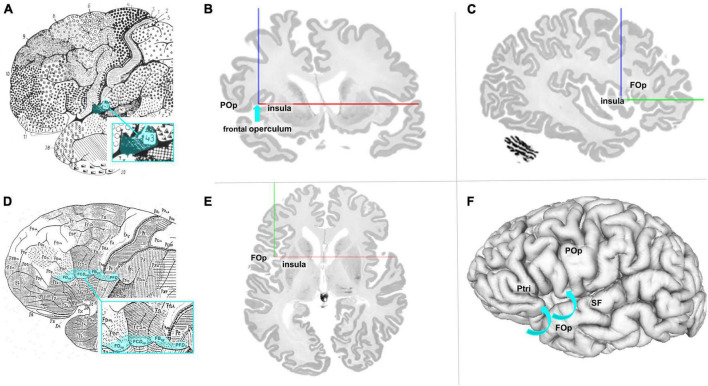
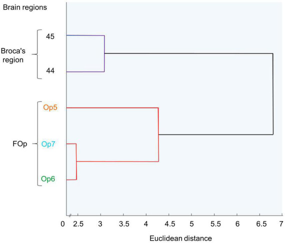
The human frontal operculum (FOp) is a brain region that covers parts of the ventral frontal cortex next to the insula. Functional imaging studies showed activations in this region in tasks related to language, somatosensory, and cognitive functions. While the precise cytoarchitectonic areas that correlate to these processes have not yet been revealed, earlier receptorarchitectonic analysis resulted in a detailed parcellation of the FOp. We complemented this analysis by a cytoarchitectonic study of a sample of ten postmortem brains and mapped the posterior FOp in serial, cell-body stained histological sections using image analysis and multivariate statistics. Three new areas were identified: Op5 represents the most posterior area, followed by Op6 and the most anterior region Op7. Areas Op5-Op7 approach the insula, up to the circular sulcus. Area 44 of Broca's region, the most ventral part of premotor area 6, and parts of the parietal operculum are dorso-laterally adjacent to Op5-Op7. The areas did not show any interhemispheric or sex differences. Three-dimensional probability maps and a maximum probability map were generated in stereotaxic space, and then used, in a first proof-of-concept-study, for functional decoding and analysis of structural and functional connectivity. Functional decoding revealed different profiles of cytoarchitectonically identified Op5-Op7. While left Op6 was active in music cognition, right Op5 was involved in chewing/swallowing and sexual processing. Both areas showed activation during the exercise of isometric force in muscles. An involvement in the coordination of flexion/extension could be shown for the right Op6. Meta-analytic connectivity modeling revealed various functional connections of the FOp areas within motor and somatosensory networks, with the most evident connection with the music/language network for Op6 left. The new cytoarchitectonic maps are part of Julich-Brain, and publicly available to serve as a basis for future analyses of structural-functional relationships in this region.
人类额盖(FOp)是一个覆盖岛叶旁腹侧额叶皮质部分区域的脑区。功能成像研究表明,在与语言、躯体感觉和认知功能相关的任务中,该区域会被激活。虽然尚未揭示与这些过程相关的精确细胞构筑区域,但早期的受体构筑分析对额盖进行了详细的分区。我们通过对10个死后大脑样本进行细胞构筑研究对这一分析进行了补充,并使用图像分析和多元统计方法,在连续的、细胞体染色的组织学切片中绘制了额盖后部的图谱。确定了三个新区域:Op5代表最后部的区域,其次是Op6和最前部的区域Op7。Op5 - Op7区域靠近岛叶,直至环状沟。布罗卡区的44区、运动前区6最腹侧部分以及顶叶盖的部分区域在背外侧与Op5 - Op7相邻。这些区域未显示出任何半球间或性别差异。在立体定向空间中生成了三维概率图谱和最大概率图谱,然后在第一个概念验证研究中用于功能解码以及结构和功能连接性分析。功能解码揭示了细胞构筑学上确定的Op5 - Op7的不同特征。左侧Op6在音乐认知中活跃,而右侧Op5参与咀嚼/吞咽和性活动处理。在肌肉进行等长力量运动时,两个区域均显示出激活。右侧Op6显示出参与屈伸协调。元分析连接性建模揭示了额盖区域在运动和躯体感觉网络内的各种功能连接,其中左侧Op6与音乐/语言网络的连接最为明显。新的细胞构筑图谱是朱利希脑图谱的一部分,可公开获取,为该区域未来结构 - 功能关系分析提供基础。